- Physical Examination
- Surgical Examination
- Ophthalmology
- Clinical Skills
- Orthopedics
- Surgery Videos
- Laparoscopy
- Pediatrics
- Funny Videos
- Cardiothoracic Surgery
- Nursing Videos
- Plastic Surgery
- Otorhinolaryngology
- Histology and Histopathology
- Neurosurgery
- Dermatology
- Pediatric Surgery
- Urology
- Dentistry
- Oncology and Cancers
- Anatomy Videos
- Health and Fitness
- Radiology
- Anaesthesia
- Physical Therapy
- Pharmacology
- Interventional Radiology
- Cardiology
- Endocrinology
- Gynecology
- Emergency Medicine
- Psychiatry and Psychology
- Childbirth Videos
- General Medical Videos
- Nephrology
- Physiology
- Diet and Food Health
- Diabetes Mellitus
- Neurology
- Women Health
- Osteoporosis
- Gastroenterology
- Pulmonology
- Hematology
- Rheumatology
- Toxicology
- Nuclear Medicine
- Infectious Diseases
- Vascular Disease
- Reproductive Health
- Burns and Wound Healing
- Other
Surgery Videos
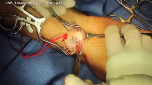
Surgery involves removing the cyst as well as part of the involved joint capsule or tendon sheath, which is considered the root of the ganglion. Even after excision, there is a small chance the ganglion will return. A ganglion cyst at the wrist is removed during a surgical procedure called excision.
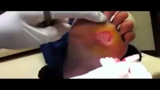
Remove a Plantar Wart from a foot Procedure
Compromise of the blood supply from microvascular disease, often in association with lack of sensation because of neuropathy, predisposes persons with diabetes mellitus to foot infections. These infections span the spectrum from simple, superficial cellulitis to chronic osteomyelitis. Diabetic foot infections typically take one of the following forms: Cellulitis Deep-skin and soft-tissue infections Acute osteomyelitis Chronic osteomyelitis Cellulitis Tender, erythematous, nonraised skin lesions are present, sometimes with lymphangitis Lymphangitis suggests group A streptococcal infection Bullae are typical of Staphylococcus aureus infection, but occasionally occur with group A streptococci
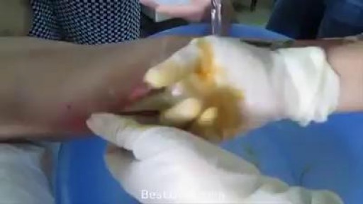
Paronychias are most often caused by common skin bacteria (most commonly staphylococci bacteria) entering the skin around the nail that has been damaged by trauma, such as nail biting, finger sucking, dishwashing, or chemical irritants. Fungal infection also can be a cause of paronychia formation and should be considered especially in people with recurrent infection. Paronychia should not be confused with herpetic whitlow, which can form tiny pustules on the finger and is caused by a virus but is not typically located at the nail edge. Herpetic whitlow is not treated with an incision and drainage and therefore needs to be distinguished from a paronychia.
Infection leg gets cleaning inside
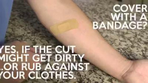
Most minor cuts you can treat yourself -- but know when to see a doctor:
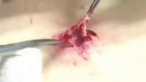
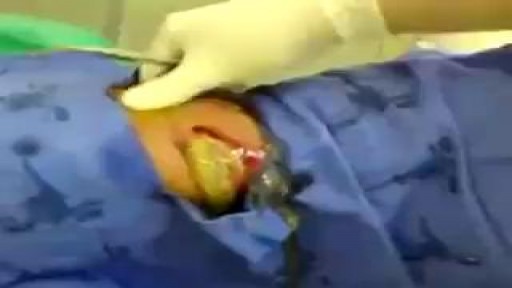
What is inside A Cyst? Watch it now
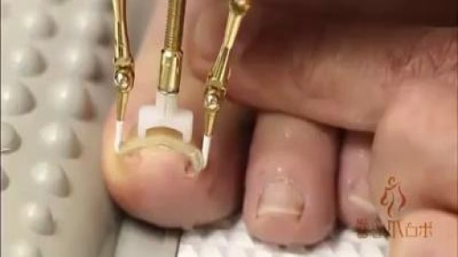
Wow! amazing tool. The disturbingly fascincating fix of ingrown toenail

The cyst was technically 46.5 pounds and her doctors call it the largest in world history. I am not sure if that is true, but it is a massive cyst
In this video we give examples of five proven techniques for popping. Viewer discretion is advised as this may not be something that all people want to see. Popping isn't for everyone!
Breast abscesses are often linked to mastitis – a condition that causes breast pain and swelling (inflammation), and usually affects women who are breastfeeding. Infections can occur during breastfeeding if bacteria enter your breast tissue, or if the milk ducts (tiny tubes) become blocked. This can cause mastitis which, if not treated, can result in an abscess forming. Women who aren't breastfeeding can also develop mastitis if bacteria enter the milk ducts through a sore or cracked nipple, or a nipple piercing. White blood cells are sent to attack the infection, which causes tissue at the site of the infection to die. This creates a small, hollow area that fills with pus (an abscess).
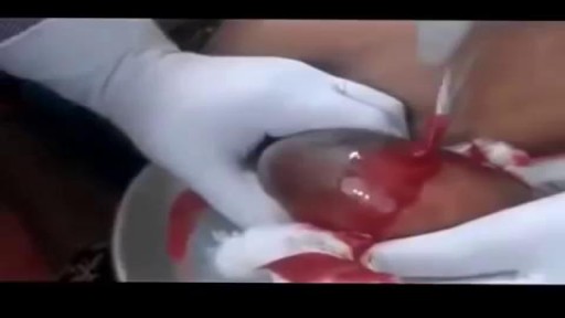
The largest amount of pus I have ever seen!!
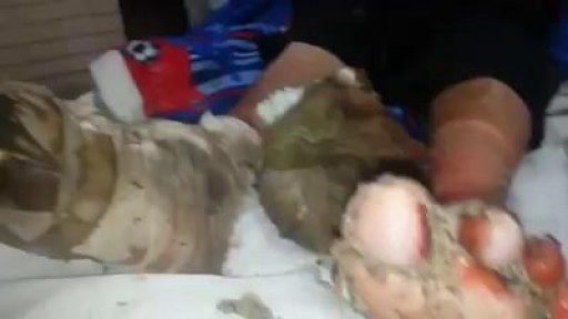
A patient suffering from Diabetic gangrene and maneged by "myiasis"

Robotic snakes help surgeons access complex anatomical locations.
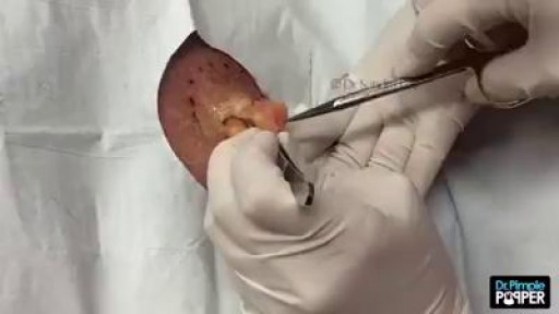
A lipoma is a growth of fat cells in a thin, fibrous capsule usually found just below the skin. Lipomas aren't cancer and don't turn into cancer. They are found most often on the torso, neck, upper thighs, upper arms, and armpits, but they can occur almost anywhere in the body. One or more lipomas may be present at the same time.
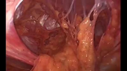
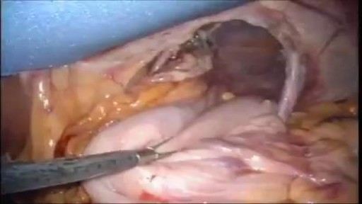
A giant abdominal wall hernia can develop from an existing ventral or incisional hernia, sometimes arising after one or more failed repair attempts. These hernias may also result from a traumatic injury where the abdomen was required to be left open and healing was delayed. In giant abdominal wall hernias, multiple loops of intestines and sometimes other abdominal organs reside within the hernia sac. The abdominal wall muscles then become conditioned to this and retract reducing the available space inside the abdomen.
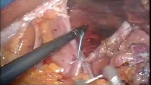
Hiatal hernias occur when contents of the abdominal cavity protrude through the esophageal hiatus of the diaphragm. Factors that contribute to the development of a hiatal hernia include an enlargement of the esophageal hiatus due to developmental defects, an increased abdominal thoracic pressure gradient, and the depletion of elastic fibers in the phrenoesophageal membrane with aging. There are four different types of hiatal hernias and management varies depending on the type. Type I, also known as a sliding hernia, is a simple displacement of the gastroesophageal junction into the thoracic cavity. The stomach remains in the abdominal cavity. This is the most common type of hiatal hernia, accounting for about 95% of all hiatal hernias. Types II-IV are classified as paraesophageal hernias. Type II occurs when the gastroesophageal junction maintains its position but the gastric fundus herniates through the diaphragmatic hiatus. Type III has both the gastroesophageal junction and the stomach herniate above the diaphragm. When more than 30% of the stomach is herniated into the thoracic cavity, it is termed a “giant” paraesophageal hernia. A patient has a type IV hernia when other organs, such as the colon, in addition to the stomach herniate above the diaphragm.
A hiatal hernia occurs when the upper part of the stomach pushes through an opening in the diaphragm and into the chest cavity. The diaphragm is the thin muscle wall that separates the chest cavity from the abdomen. The opening in the diaphragm is where the esophagus and stomach join.
3D animation video of Varicose Veins Sclerotherapy Treatment
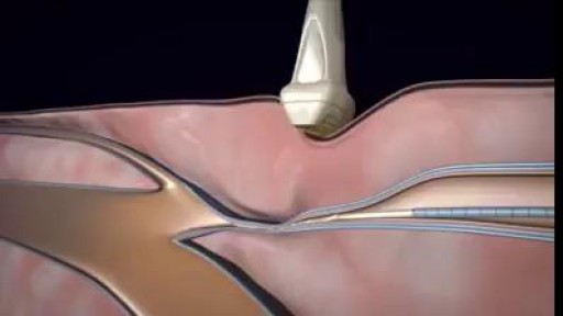
Surgical Repair of Pectus Excavatum. Pectus excavatum is a condition in which a person's breastbone is sunken into his or her chest.
