Histology and Histopathology
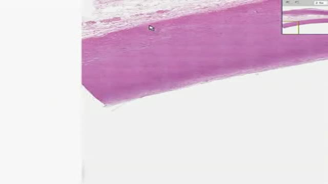
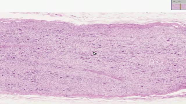
Histology of Aorta

Histology of Areolar Connective Tissue
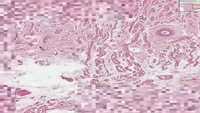
Histology of Tongue
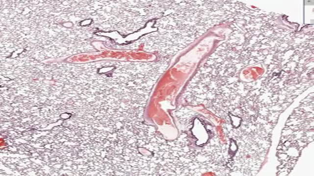
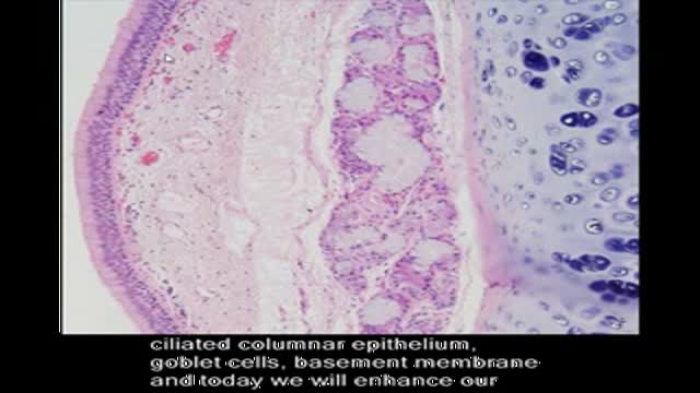
Histology of Lung
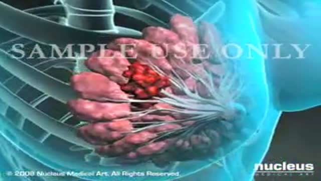
This 3D medical animation shows several methods of breast tissue biopsy procedures including:
- Needle biopsy,
- Stereotactic core biopsy
- Ultrasound-guided core biopsy - - Surgical biopsy
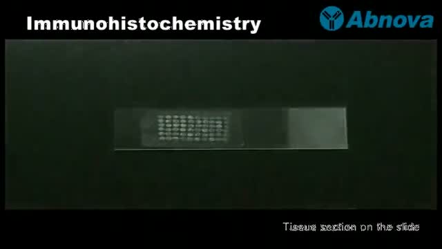
Immunohistochemistry or IHC refers to the process of detecting antigens (e.g., proteins) in cells of a tissue section by exploiting the principle of antibodies binding specifically to antigens in biological tissues.[1] IHC takes its name from the roots "immuno," in reference to antibodies used in the procedure, and "histo," meaning tissue (compare to immunocytochemistry). Immunohistochemical staining is widely used in the diagnosis of abnormal cells such as those found in cancerous tumors. Specific molecular markers are characteristic of particular cellular events such as proliferation or cell death (apoptosis). IHC is also widely used in basic research to understand the distribution and localization of biomarkers and differentially expressed proteins in different parts of a biological tissue. Visualising an antibody-antigen interaction can be accomplished in a number of ways. In the most common instance, an antibody is conjugated to an enzyme, such as peroxidase, that can catalyse a colour-producing reaction. Alternatively, the antibody can also be tagged to a fluorophore, such as fluorescein or rhodamine
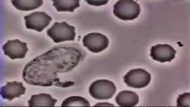
A very interesting video showing how white blood cells (Neutrophil) are chasing bacteria (Diplococci). It also shows how the white blood cell engulf the bacteria. This is a real video.
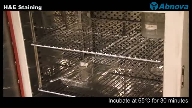
H&E stain is a popular staining method in histology. Its a combination of two dyes: the basic dye (hematoxylin) and the alcohol-based dye (eosin). In an H&E stain you will usually see both eosinophilia and basophilia: the nuclei of cells basophilic (blue), while eosinophilia is typical of cytoplasmic constituents (pink). Xylene, alcohols, distilled water are also required.
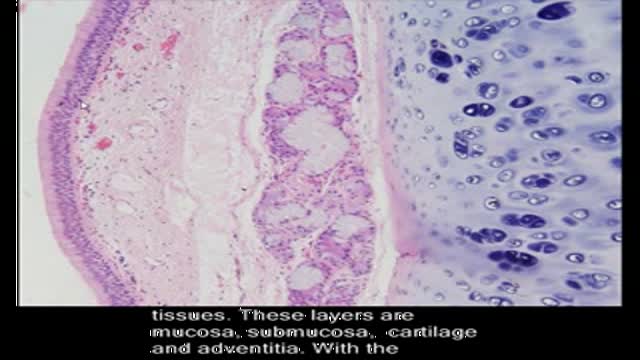
the short video will describe four layers of connective tissue. Please see disclaimer on my website. www.academyofprofessionals.com
A few words on connective tissue. Please see disclaimer on my website www.academyofprofessionals.com
a video showing the technique of Shave and Punch Skin Biopsies nique of
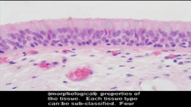
The video will describe epithelium. Please see disclaimer on my website. www.academyofprofessionals.com
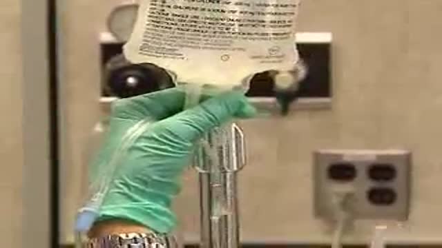
Intravenous line and cannula insertion
Histology of Sympathetic Ganglion
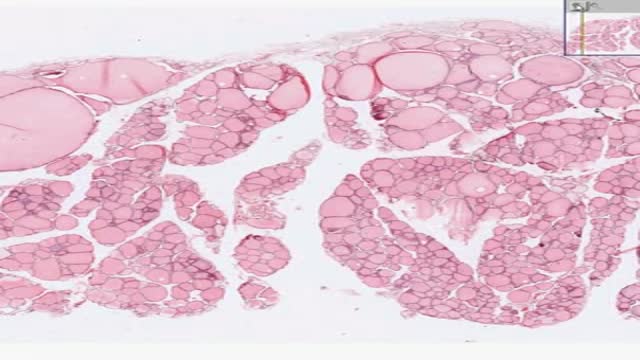
Histology of the Thyroid gland
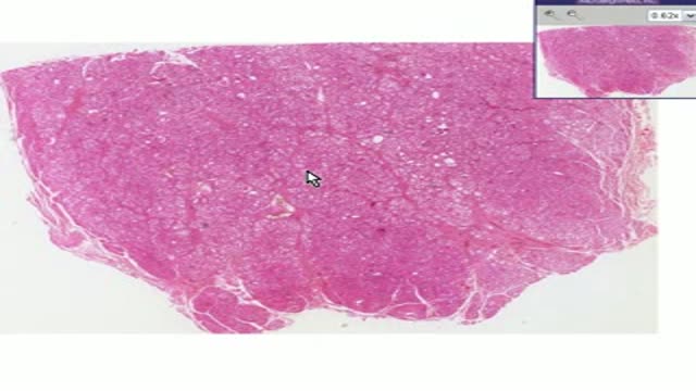
Histopathology of Graves Disease
