Anatomy Videos

Microsoft HoloLens. Medical Education

For education, Microsoft HoloLens will help make incredible leaps forward in productivity, collaboration, and innovation. See how Microsoft HoloLens transforms the way we teach anatomy and our understanding of the human body as we help to prepare the next generation of doctors.
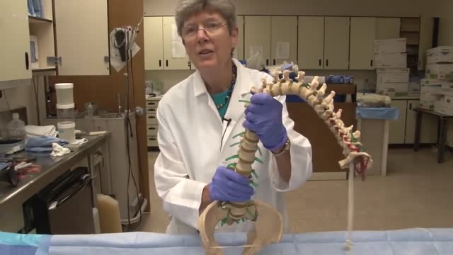
The spinal cord is a long, thin, tubular bundle of nervous tissue and support cells that extends from the medulla oblongata in the brainstem to the lumbar region of the vertebral column. The brain and spinal cord together make up the central nervous system (CNS).
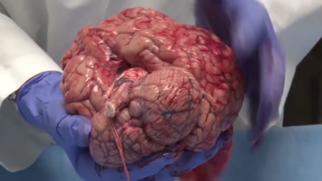
The human brain is the command center for the human nervous system. It receives input from the sensory organs and sends output to the muscles. The human brain has the same basic structure as other mammal brains, but is larger in relation to body size than any other brains.
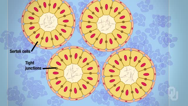
The male reproductive system includes the scrotum, testes, spermatic ducts, sex glands, and penis. These organs work together to produce sperm, the male gamete, and the other components of semen.
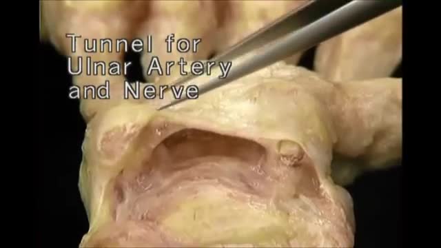
Hand Anatomy

Clinical Anatomy Lecture Illustrate The Anatomy Of The Abdominal Wall
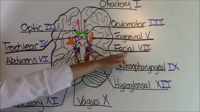
Cranial Nerves Mnemonic
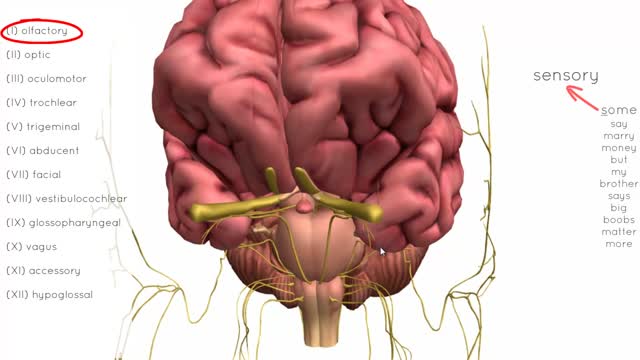
There are twelve cranial nerves in total. The olfactory nerve (CN I) and optic nerve (CN II) originate from the cerebrum. Cranial nerves III – XII arise from the brain stem (Figure 1). They can arise from a specific part of the brain stem (midbrain, pons or medulla), or from a junction between two parts: Midbrain – the trochlear nerve (IV) comes from the posterior side of the midbrain. It has the longest intracranial length of all the cranial nerves. Midbrain-pontine junction – oculomotor (III). Pons – trigeminal (V). Pontine-medulla junction – abducens, facial, vestibulocochlear (VI-VIII). Medulla Oblongata – posterior to the olive: glossopharyngeal, vagus, accessory (IX-XI). Anterior to the olive: hypoglossal (XII). The cranial nerves are numbered by their loca
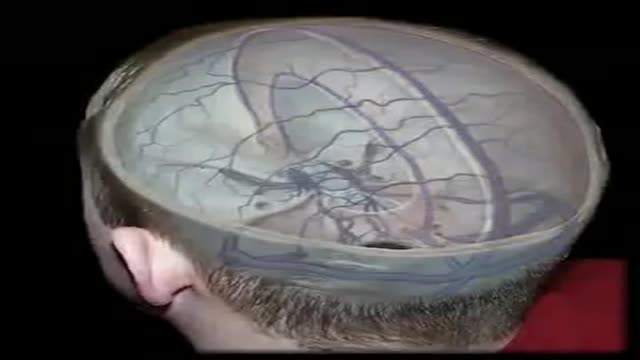
The dural venous sinuses are spaces between the endosteal and meningeal layers of the dura. They contain venous blood that originates for the most part from the brain or cranial cavity. The sinuses contain an endothelial lining that is continuous into the veins that are connected to them.

The superior sagittal sinus (also known as the superior longitudinal sinus), within the human head, is an unpaired area along the attached margin of falx cerebri. It allows blood to drain from the lateral aspects of anterior cerebral hemispheres to the confluence of sinuses.
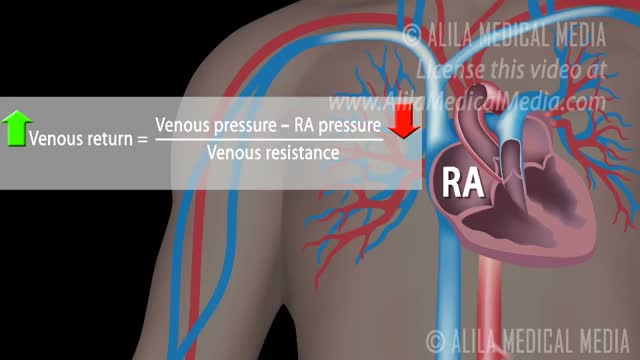
How Respiratory Pump Affects Venous Return
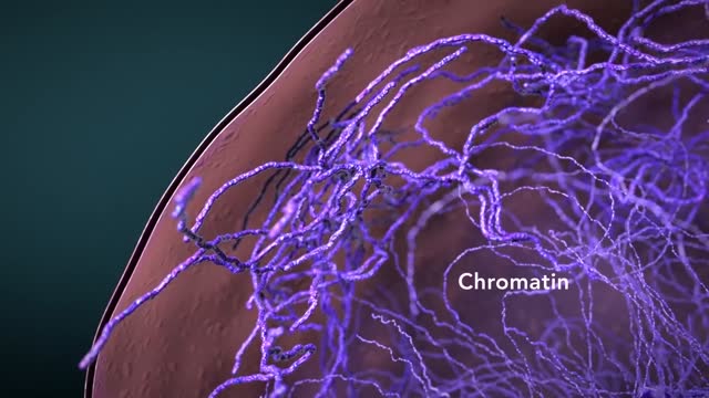
Cytoplasmic organelles are "little organs" that are suspended in the cytoplasm of the cell. Each type of organelle has a definite structure and a specific role in the function of the cell. Examples of cytoplasmic organelles are mitochondrion, ribosomes, endoplasmic reticulum, golgi apparatus, and lysosomes.
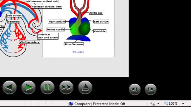
Embryonic cardiovascular system. ... The human arterial and venous systems develop from different embryonic areas. Aortic Arches. The aortic arches—or pharyngeal arch arteries—are a series of six, paired, embryological vascular structures that give rise to several major arteries .
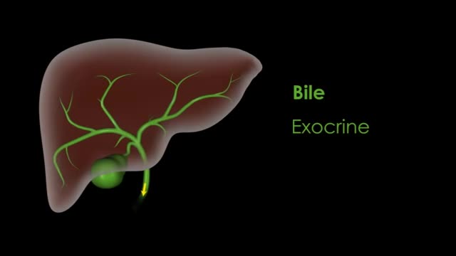
liver also detoxifies chemicals and metabolizes drugs. As it does so, the liver secretes bile that ends up back in the intestines. The liver also makes proteins important for blood clotting and other functions.
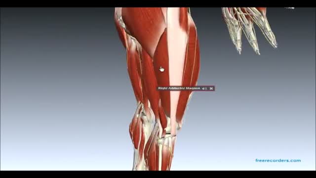
Muscles and Nerves of Lower Limb
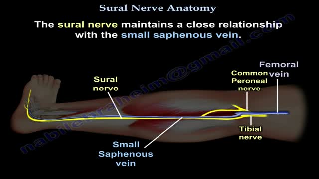
Nerves are the organs that make up the peripheral nervous system (PNS). They serve as information pipelines that allow the brain and spinal cord to communicate with other tissues and organs. Inside the nerves are the axon processes of sensory and motor neurons (nerve cells).
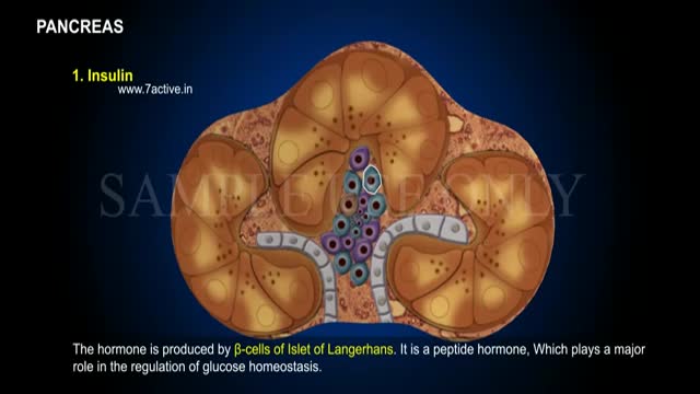
Enzymes, or digestive juices, produced by the pancreas are secreted into the small intestine to further break down food after it has left the stomach. The gland also produces the hormone insulin and secretes it into the bloodstream in order to regulate the body's glucose or sugar level.
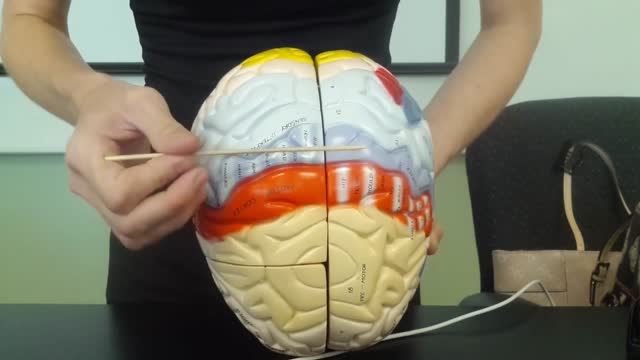
The brain is that part of the CNS contained within the cranial cavity (figure 13.1). It is the control center for many of the body's functions. The brain is much like a complex central computer but with additional functions that no computer can as yet match. Indeed, one goal in computer technology is to make computers that can function more like the human brain. The brain consists of the brainstem, the cerebellum, the diencephalon, and the cerebrum (table 13.1). The brainstem includes the medulla oblongata, pons, midbrain, and reticular formation. The structure of the brain is described in this chapter. Its functions are primarily discussed in chapter 14. Twelve pairs of cranial nerves, which are part of the PNS, arise directly from the brain. Two pairs arise from the cerebrum, nine pairs arise from the brainstem, and one pair arises from the spinal cord.
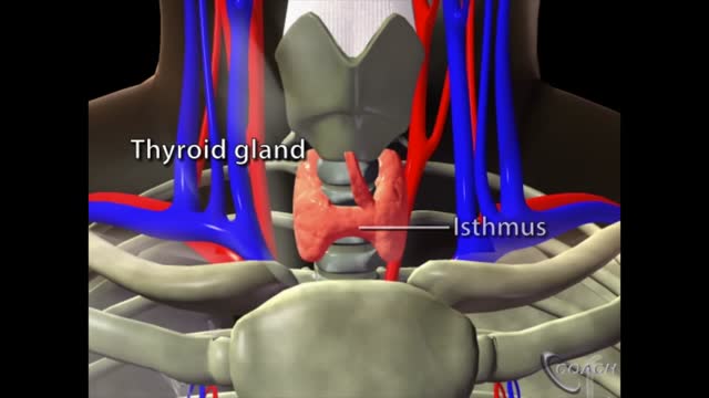
The thyroid is a butterfly-shaped gland that sits low on the front of the neck. Your thyroid lies below your Adam’s apple, along the front of the windpipe. The thyroid has two side lobes, connected by a bridge (isthmus) in the middle. When the thyroid is its normal size, you can’t feel it.
