- Physical Examination
- Surgical Examination
- Ophthalmology
- Clinical Skills
- Orthopedics
- Surgery Videos
- Laparoscopy
- Pediatrics
- Funny Videos
- Cardiothoracic Surgery
- Nursing Videos
- Plastic Surgery
- Otorhinolaryngology
- Histology and Histopathology
- Neurosurgery
- Dermatology
- Pediatric Surgery
- Urology
- Dentistry
- Oncology and Cancers
- Anatomy Videos
- Health and Fitness
- Radiology
- Anaesthesia
- Physical Therapy
- Pharmacology
- Interventional Radiology
- Cardiology
- Endocrinology
- Gynecology
- Emergency Medicine
- Psychiatry and Psychology
- Childbirth Videos
- General Medical Videos
- Nephrology
- Physiology
- Diet and Food Health
- Diabetes Mellitus
- Neurology
- Women Health
- Osteoporosis
- Gastroenterology
- Pulmonology
- Hematology
- Rheumatology
- Toxicology
- Nuclear Medicine
- Infectious Diseases
- Vascular Disease
- Reproductive Health
- Burns and Wound Healing
- Other
Radiology

Head CT Interpretation Made Easy


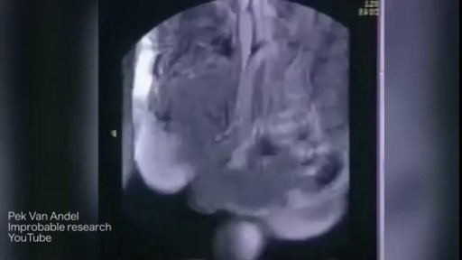
Watch that video of MRI Scans Human Body Internal Organs During Sex
Pregnancy ultrasounds are performed mainly using transabdominal ultrasound. For many women, especially after 8 weeks gestation, sufficient information about the baby may be obtained with transabdominal ultrasound only. However, in the early pregnancy, the developing embryo is very small (at 6 weeks gestation, the baby is only 5-9mm long) and a transvaginal ultrasound may be required to get a better image of the baby. Transvaginal ultrasound is safe and commonly performed during all stages of pregnancy, including the first trimester. It will not harm you or your baby.
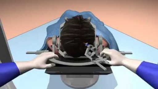
MRI-guided laser ablation for minimal invasive Neurosurgery.
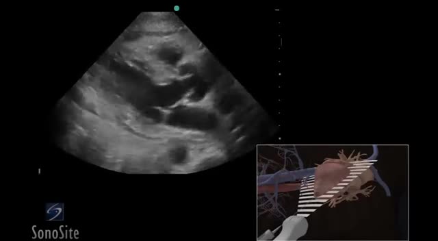
Ultrasound or ultrasonography is a medical imaging technique that uses high frequency sound waves and their echoes. The technique is similar to the echolocation used by bats, whales and dolphins, as well as SONAR used by submarines. In ultrasound, the following events happen: The ultrasound machine transmits high-frequency (1 to 5 megahertz) sound pulses into your body using a probe. The sound waves travel into your body and hit a boundary between tissues (e.g. between fluid and soft tissue, soft tissue and bone). Some of the sound waves get reflected back to the probe, while some travel on further until they reach another boundary and get reflected. The reflected waves are picked up by the probe and relayed to the machine. The machine calculates the distance from the probe to the tissue or organ (boundaries) using the speed of sound in tissue (5,005 ft/s or1,540 m/s) and the time of the each echo's return (usually on the order of millionths of a second). The machine displays the distances and intensities of the echoes on the screen, forming a two dimensional image like the one shown below.

This upgraded ultrasound device allows you to see your organs with the help of augmented reality glasses

The diffuse lung diseases tend to cause infiltrative opacification in the periphery of the lung. As the name of the group of diseases suggests, they are diffuse. While the consolidation or ground-glass change is usually bilateral, it may be localised, e.g. radiation pneumonitis.
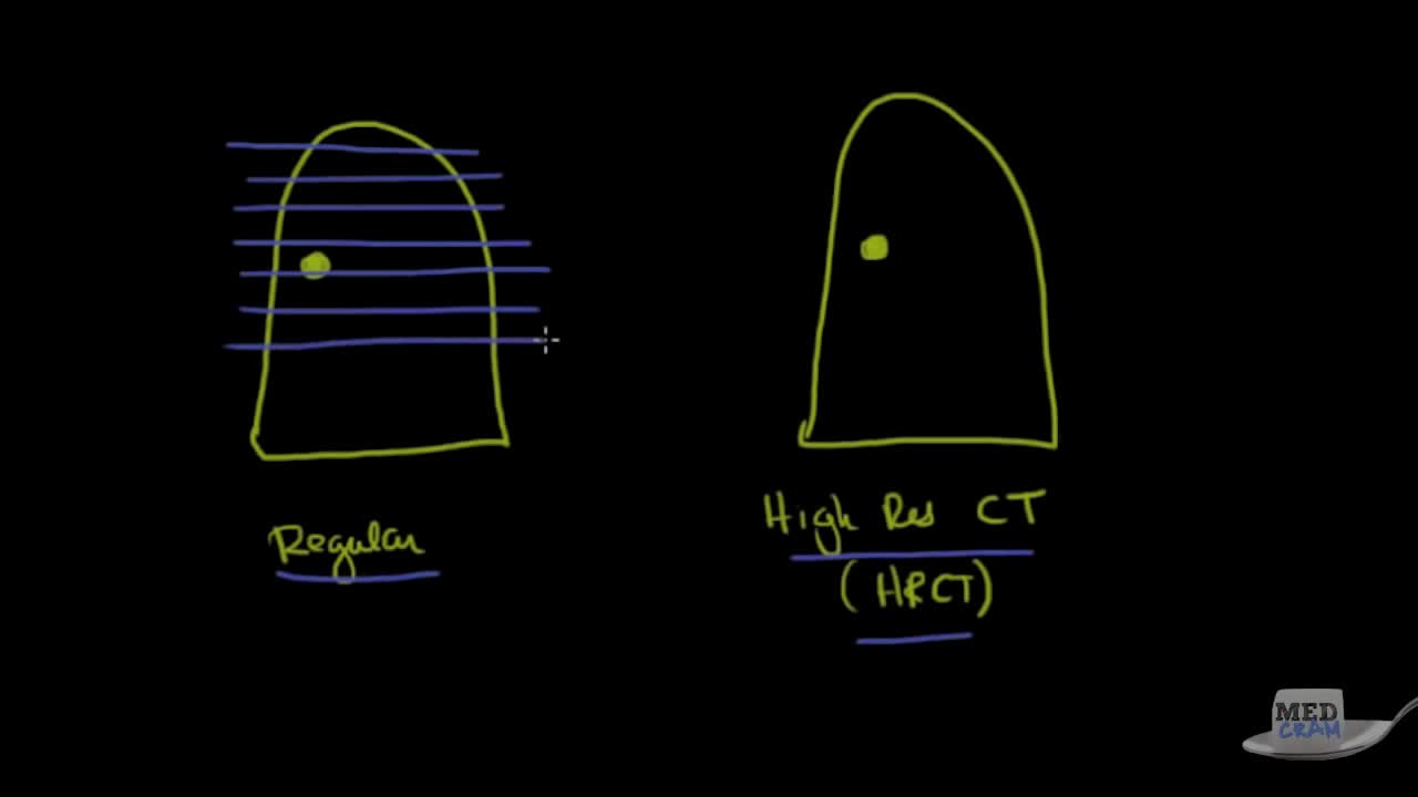
Understand Chest CT (Computed Tomography) scans with this clear explanation
Using 3D animations we have come up with a new way of demonstrating how to perform portable ultrasound examinations

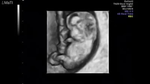
MRI Shows Twins Fighting in the Womb

Expand Section. Pulmonary edema is often caused by congestive heart failure. When the heart is not able to pump efficiently, blood can back up into the veins that take blood through the lungs. As the pressure in these blood vessels increases, fluid is pushed into the air spaces (alveoli) in the lungs.
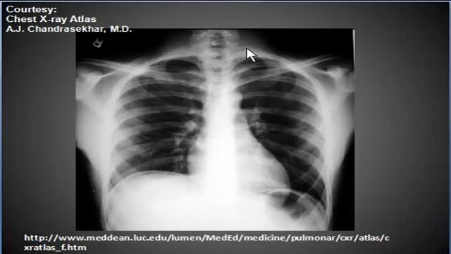
Chest x-ray, pneumoperitonuem, air under diaphragms
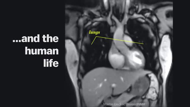
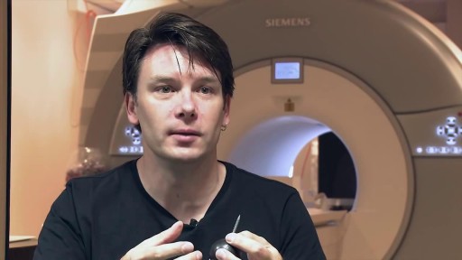
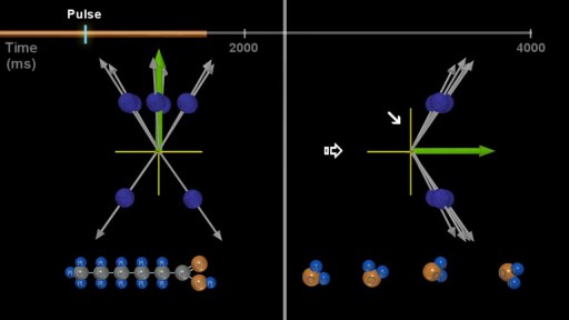
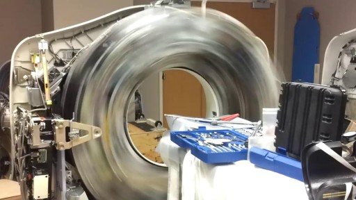
Magnetic Resonance Imaging (MRI) "sees" inside the body by mapping the position of water molecules, which exist at different densities in different types of tissue. Watch the video above for a sample of some impressive MRI images of the human body in action.
The human body as seen with MRI and X-RAY

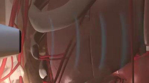
This tiny camera can capture images inside the brain.
