- Physical Examination
- Surgical Examination
- Ophthalmology
- Clinical Skills
- Orthopedics
- Surgery Videos
- Laparoscopy
- Pediatrics
- Funny Videos
- Cardiothoracic Surgery
- Nursing Videos
- Plastic Surgery
- Otorhinolaryngology
- Histology and Histopathology
- Neurosurgery
- Dermatology
- Pediatric Surgery
- Urology
- Dentistry
- Oncology and Cancers
- Anatomy Videos
- Health and Fitness
- Radiology
- Anaesthesia
- Physical Therapy
- Pharmacology
- Interventional Radiology
- Cardiology
- Endocrinology
- Gynecology
- Emergency Medicine
- Psychiatry and Psychology
- Childbirth Videos
- General Medical Videos
- Nephrology
- Physiology
- Diet and Food Health
- Diabetes Mellitus
- Neurology
- Women Health
- Osteoporosis
- Gastroenterology
- Pulmonology
- Hematology
- Rheumatology
- Toxicology
- Nuclear Medicine
- Infectious Diseases
- Vascular Disease
- Reproductive Health
- Burns and Wound Healing
- Other
Radiology
Many children receive MRIs at the hospital, and it can often be a scary experience if they are unprepared or don't know what to expect.
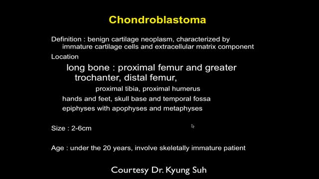
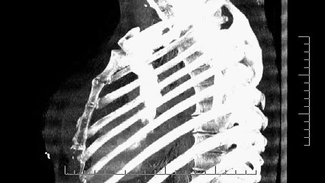
MRI of Bone Tumor

Anatomy of Love
If you have multiple sclerosis (MS), you probably had several tests done before you received your diagnosis. There isn’t one test to diagnosis MS, so testing can vary. Doctors can use neurological exams, information about previous symptoms, blood tests, and spinal fluid tests. A magnetic resonance imaging (MRI) scan isn’t used to diagnose MS but rather to rule out other diseases. A diagnosis of MS requires more information than what a scan alone can give. By looking at more than one test or exam result, doctors can get a clearer picture of what’s going on in your body.
MRI Exam Procedure
Magnetic resonance imaging (MRI) can be an important tool in the diagnosis of multiple sclerosis (MS). MRI can also be used to monitor the progression of the disease in people living with MS. How does it work? MRI uses very strong magnets, radio signals, and computer software to take 3-dimensional pictures of the inside of the body. Will I need contrast material? Maybe. Contrast material is a substance that temporarily changes the way imaging tools interact with the body. They are often used to visualize certain types of MS disease activity on the MRI. If your doctor thinks your scan requires this contrast material, you may get an injection before you get in the MRI machine. How long will it take? The time may vary based on the type of MRI. Be sure to discuss with your doctor in advance so he or she can provide you with exact timing. But don’t worry, you won’t have to stay still the whole time. The technician will let you know when they’re starting a new image.
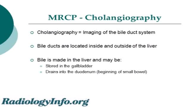
An MRCP scan is a scan that uses magnetic resonance imaging (MRI) to produce pictures of the liver, bile ducts, gallbladder and pancreas. Note: the information below is a general guide only. The arrangements,and the way tests are performed, may vary between different hospitals.

This video is a simplified tutorial to teach how to read and understand chaest x-rays. It is for beginners
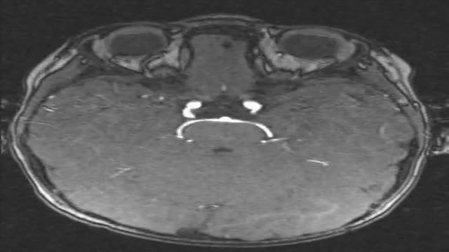
2 year old boy with chronic sinusitis, headache, vertigo problem, decreased vision and hearing. Repeated lung infections. Can you see it? Pay also special attention to the ears.

35 year old women with breathing difficulties for 6 months and feels like fluid is leaking down her front and back. Pain in thorax, lower back and pelvic. Weight loss. Was exposed to mold for a 2 years. Has a dog witch has persistent worm infection. Also been traveling out of the country.
35 year old women with breathing difficulties for 6 months and feels like fluid is leaking down her front and back. Was exposed to mold for a 2 years. Has a dog witch has persistent worm infection. Breast implants 10 years ago.

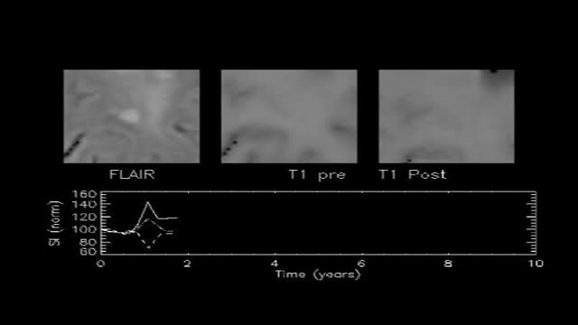
MRI of Fetal Brain Development
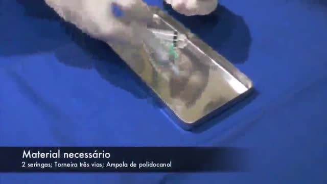
Creating polidocanol foam
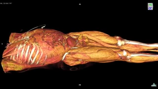
Whole Body CT scan with contrast media HD
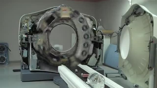
CT Scanner 64 slice Inside HD

A Bone scan or bone scintigraphy is a nuclear scanning test to find certain abnormalities in bone which are triggering the bone's attempts to heal. It is primarily used to help diagnose a number of conditions relating to bones, including: cancer of the bone or cancers that have spread (metastasized) to the bone, locating some sources of bone inflammation (e.g. bone pain such as lower back pain due to a fracture), the diagnosis of fractures that may not be visible in traditional X-ray images, and the detection of damage to bones due to certain infections and other problems.
Nuclear medicine bone scans are one of a number of methods of bone imaging, all of which are used to visually detect bone abnormalities. Such imaging studies include magnetic resonance imaging (MRI), X-ray computed tomography (CT) and in the case of 'bone scans' nuclear medicine. However, a nuclear bone scan is a functional test, which means it measures an aspect of bone metabolism, which most other imaging techniques cannot. The nuclear bone scan competes with the FDG-PET scan in seeing abnormal metabolism in bones, but it is considerably less expensive.
Nuclear bone scans are not to be confused with the completely different test often termed a "bone density scan," DEXA or DXA, which is a low exposure X-ray test measuring bone density to look for osteoporosis and other diseases where bones lose mass, without any bone re-building activity. The nuclear medicine scan technique is sensitive to areas of unusual bone re-building activity because the radiopharmaceutical is taken up by osteoblast cells which build bone. The technique therefore is sensitive to fractures and bone reaction to infections and bone tumors, including tumor metastases to bones, because all these pathologies trigger bone osteoblast activity. The bone scan is not sensitive to osteoporosis or multiple myeloma in bones, and therefore other techniques must be used to assess bone abnormalities from these diseases.
Identify the anatomy and explain the physiology of the scrotum on diagrams and sonograms.
Describe and demonstrate the protocol for sonographic scanning of the scrotum.
Identify and describe sonographic images of congenital abnormalities of the scrotum.
Identify and describe sonographic images of pathologies of the scrotum.
Identify and describe sonographic images of extratesticular disease processes.
Identify the anatomy and explain the physiology of the prostate on diagrams and sonograms.
Describe and demonstrate the protocol for transabdominal and endorectal sonographic scanning of the prostate.
Identify and describe sonographic images of benign and malignant pathologies of the prostate, including benign hyperplasia, prostatitis, carcinoma, and calculi.
Explain the technique for prostate biopsy.
Define the criteria for an ultrasound appearance of prostate tumor staging.
Explain the technique for radiation seed implantation.
Explain the Patient Privacy Rule (HIPAA) and Patient Safety Act (see reference).
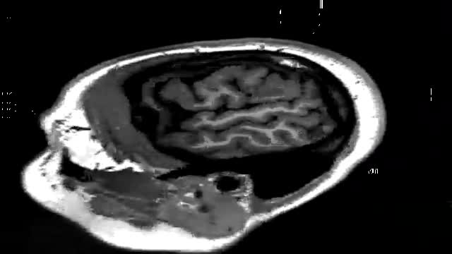
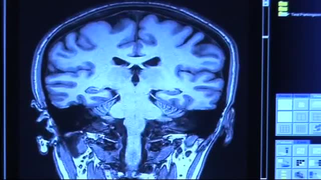
An animated video showing an MRI of the brain

MPG Video for purpose

n,n,n,mm
