Cardiology
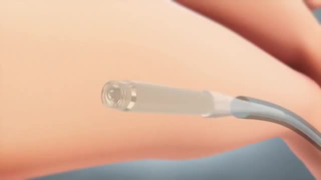
This tiny wireless pacemaker can be inserted into the body via a catheter instead of invasive surgery.

Restrictive cardiomyopathy (RCM) is a rare form of heart muscle disease that is characterized by restrictive filling of the ventricles. In this disease the contractile function (squeeze) of the heart and wall thicknesses are usually normal, but the relaxation or filling phase of the heart is very abnormal.
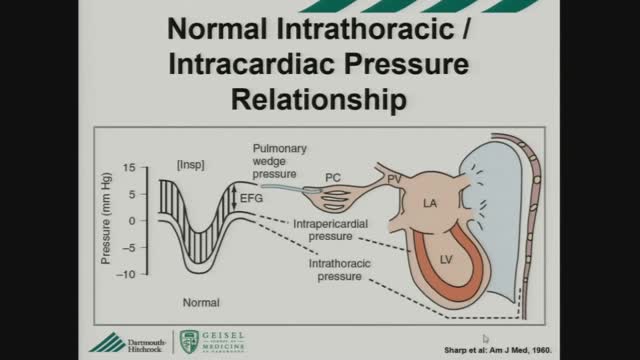
Constrictive pericarditis is the result of scarring and consequent loss of the normal elasticity of the pericardial sac. This leads to impairment of ventricular filling in mid and late diastole. As a result, the majority of ventricular filling occurs rapidly in early diastole and the ventricular volume does not increase after the end of the early filling period. Restrictive cardiomyopathy is characterized by a nondilated rigid ventricle, resulting in severe diastolic dysfunction and restrictive filling that produces hemodynamic changes similar to those in constrictive pericarditis. Constrictive pericarditis and restrictive cardiomyopathy both lead to diastolic heart failure with normal (or near normal) systolic function, and characteristically abnormal ventricular filling that results in similar clinical and hemodynamic features. However, because of their markedly different treatments, differentiating between the two conditions is critical. In some patients, the correct diagnosis may be readily suggested from the history or routine diagnostic testing. In others, however, this differentiation cannot be diagnosed before biopsy or even surgical exploration.
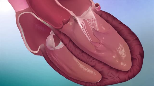
Hypertrophic cardiomyopathy (HCM) is very common and can affect people of any age. It affects men and women equally. It is a common cause of sudden cardiac arrest in young people, including young athletes. Hypertrophic cardiomyopathy occurs if heart muscle cells enlarge and cause the walls of the ventricles (usually the left ventricle) to thicken. The ventricle size often remains normal, but the thickening may block blood flow out of the ventricle. If this happens, the condition is called obstructive hypertrophic cardiomyopathy. Sometimes the septum, the wall that divides the left and right sides of the heart, thickens and bulges into the left ventricle. This can block blood flow out of the left ventricle. Then the ventricle must work hard to pump blood. Symptoms can include chest pain, dizziness, shortness of breath, or fainting. Hypertrophic cardiomyopathy also can affect the heart's mitral valve, causing blood to leak backward through the valve. Sometimes, the thickened heart muscle doesn't block blood flow out of the left ventricle. This is referred to as non-obstructive hypertrophic cardiomyopathy. The entire ventricle may thicken, or the thickening may happen only at the bottom of the heart. The right ventricle also may be affected. In both obstructive and non-obstructive HCM, the thickened muscle makes the inside of the left ventricle smaller, so it holds less blood. The walls of the ventricle may stiffen, and as a result, the ventricle is less able to relax and fill with blood.
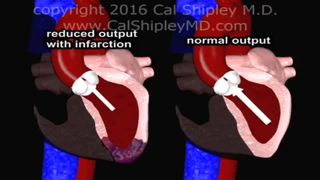
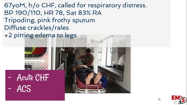
Cardiogenic shock is a condition in which your heart suddenly can't pump enough blood to meet your body's needs. The condition is most often caused by a severe heart attack. Cardiogenic shock is rare, but it's often fatal if not treated immediately. If treated immediately, about half the people who develop the condition survive.
High blood pressure and high cholesterol are the most common causes of these spasms. Approximately 2 percent of people with angina, or chest pain and pressure, experience coronary artery spasms. Coronary artery spasms can also occur in people who have atherosclerosis.
Chronic angina is a prevalent manifestation of cardiovascular disease and is most commonly due to insufficient oxygen supply from fixed epicardial lesions in the coronary arteries.
Hypertensive urgency must be distinguished from hypertensive emergency. Urgency is defined as severely elevated blood pressure (ie, systolic >220 mm Hg or diastolic >120 mm Hg) with no evidence of target organ damage.
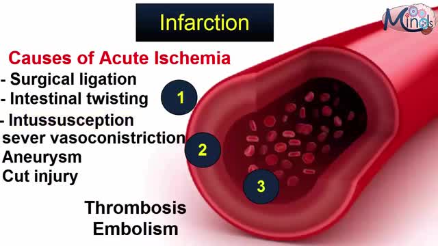
The occurrence and extent of cerebral infarction is determined by three basic factors: i) site of arterial occlusion, ii) the rapidity of arterial occlusion, and iii) the presence or absence of collateral circulation. Grossly, infarcts are usually divided into pale (non-hemorrhagic) and hemorrhagic types. Infarcts evolve over time, thus their gross appearance gives a clue to when they occurred. The temporal evolution of an infarct occurs in three stages: i) acute (1 day – 1 week) – the involved area is soft and edematous and there is a blurring of anatomic detail; ii) subacute (1 week – 1 month) – there is obvious tissue destruction and liquefactive necrosis of the involved brain; iii) chronic (>1 month) – the damaged tissue has been phagocytized and there is cavition with surrounding gliosis. Microscopically there is also a temporal evolution of cerebral infarcts. During the earliest phase of infarction (0-48 hours) chromatolysis and swollen eosinophilic neurons are seen. Neuronal cell necrosis and an acute inflammatory response are usually seen from 24-72 hours. This response is typically followed by an influx of mononuclear cells which begin to phagocytize necrotic debris (3-5 days). From 1-2 weeks after the infarct there is vascular proliferation and reactive astrocytosis. Over time (>1 month) the necrotic tissue will be completely removed and a cystic cavity surrounded by a glial scar will be formed.
Take regular breaks. If you are in a prolonged standing position or a prolonged sitting position, take regular breaks and move your arms or legs. Take a short walk, do some leg or arm exercises, on the spot walking/running, or take a walk outside the workplace. Get your circulation moving to your extremities.
Claudication, which is defined as reproducible ischemic muscle pain, is one of the most common manifestations of peripheral arterial occlusive disease (PAOD) caused by atherosclerosis. Claudication occurs during physical activity and is relieved after a short rest. Pain develops because of inadequate blood flow.
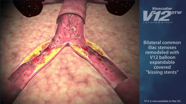
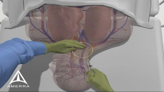
Indications for endovascular repair of the iliac artery are: Stenosis or (short-segment) occlusion of iliac artery (TASC type A and B, TASC C lesions are controversial) with ipsilateral lower extremity ischemia (lifestyle-limiting, progressive claudication, rest pain, gangrene). Patients with asymptomatic aneurysm greater than 4 cm in diameter. An iliac aneurysm which has also increased in size by 0.5 cm in last six months. Symptomatic iliac artery aneurysms mandate endovascular (or open) repair regardless of size. Patients with long occluded lesions/poor run-off/acute limb ischemia are poor endovascular candidates.
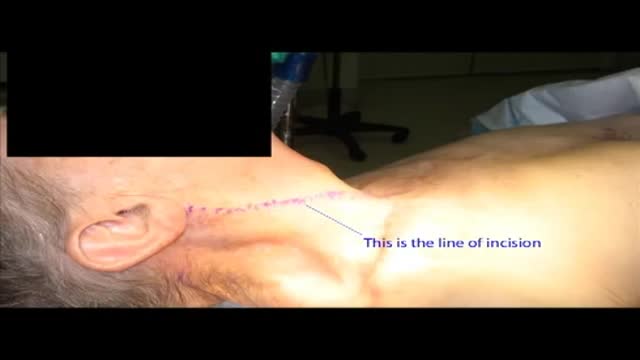
Carotid artery stenosis can be caused by cholesterol build-up in the blood vessels (atherosclerosis). Blood clots can form in this area and travel up to the brain. This condition may be present for a long time before symptoms appear. When symptoms do occur, stroke or brief stroke-like attacks are common. If this condition is discovered as a result of a stroke or stroke-like attack, cholesterol lowering medications and blood thinners may be used to improve blood flow to the brain. If the degree of narrowing is severe, surgery may be needed to open the blood vessel.
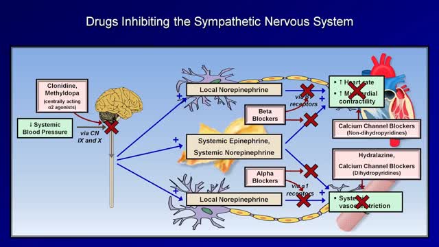
Medications to treat high blood pressure Thiazide diuretics. ... Beta blockers. ... Angiotensin-converting enzyme (ACE) inhibitors. ... Angiotensin II receptor blockers (ARBs). ... Calcium channel blockers. ... Renin inhibitors
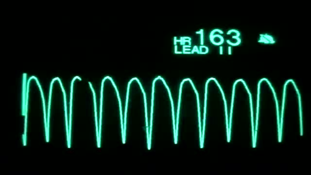
On the rhythm strip, the QRS might be somewhat taller or wider. One commonly seen type of polymorphic ventricular tachycardia is torsades de pointes. Torsades and other polymorphic VT are advanced rhythms which require additional expertise and expert consultation is advised.
If you or someone you love has atrial fibrillation, learn more about what AFib is, why treatment can save lives, and what you can do to reach your goals, lower your risks and live a healthy life.
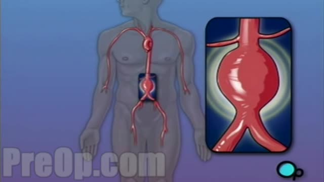
For this surgery, your doctor makes a large incision in the abdomen to expose the aorta. Once he or she has opened the abdomen, a graft can be used to repair the aneurysm. Open repair remains the standard procedure for an abdominal aortic aneurysm repair. Endovascular aneurysm repair (EVAR).
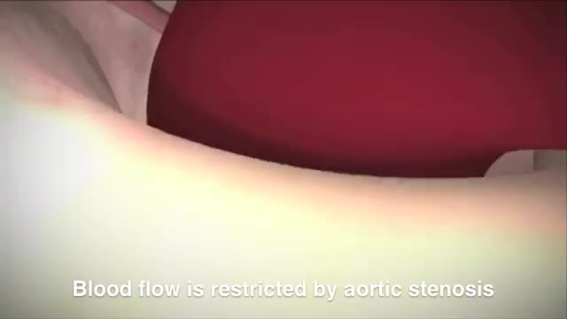
This minimally invasive surgical procedure repairs the valve without removing the old, damaged valve. Instead, it wedges a replacement valve into the aortic valve’s place. The surgery may be called a transcatheter aortic valve replacement (TAVR) or transcatheter aortic valve implantation (TAVI).
Catheters can be placed in veins in the neck (internal jugular vein), chest (subclavian vein or axillary vein), groin (femoral vein), or through veins in the arms (also known as a PICC line, or peripherally inserted central catheters).
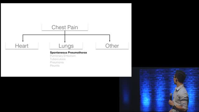
Chest pain is a frequent complaint of patients seeking urgent medical assistance, and accounts for an estimated 2-4 per cent of all A&E visits in the UK (Becker, 2000). Generally, acute chest pain should be considered cardiovascular in origin until proven otherwise and it is common in clinical practice to err on the conservative or ‘safe’ side when evaluating people with chest pain. Individuals with suspected ischaemic chest pain must be evaluated rapidly for several reasons: - Myocardial ischaemia, if prolonged and severe, can cause myocardial infarction (necrosis); - Treatment strategies that achieve myocardial salvage (thrombolytic therapy or primary coronary angioplasty) are available for patients with acute coronary syndromes and these treatments reduce morbidity and mortality;
