- Physical Examination
- Surgical Examination
- Ophthalmology
- Clinical Skills
- Orthopedics
- Surgery Videos
- Laparoscopy
- Pediatrics
- Funny Videos
- Cardiothoracic Surgery
- Nursing Videos
- Plastic Surgery
- Otorhinolaryngology
- Histology and Histopathology
- Neurosurgery
- Dermatology
- Pediatric Surgery
- Urology
- Dentistry
- Oncology and Cancers
- Anatomy Videos
- Health and Fitness
- Radiology
- Anaesthesia
- Physical Therapy
- Pharmacology
- Interventional Radiology
- Cardiology
- Endocrinology
- Gynecology
- Emergency Medicine
- Psychiatry and Psychology
- Childbirth Videos
- General Medical Videos
- Nephrology
- Physiology
- Diet and Food Health
- Diabetes Mellitus
- Neurology
- Women Health
- Osteoporosis
- Gastroenterology
- Pulmonology
- Hematology
- Rheumatology
- Toxicology
- Nuclear Medicine
- Infectious Diseases
- Vascular Disease
- Reproductive Health
- Burns and Wound Healing
- Other
Physical Examination

a video of abdominal physical examination including all the required items:
-Inspection
-Palpation
-Percussion
-Auscultation

Breast cancer is a malignant tumor that develops from the cells of
the breast. It is the most common type of cancer among women in
the United States. It is most often curable when found early. The
normal breast consists of three main components: the lobules
(milk-producing glands), the ducts (thin tubes that connect the
lobules to the nipple) and the stroma (fatty tissue and ligaments
surrounding the ducts and lobules, blood vessels, and lymphatic
vessels). About 80% of breast cancers start in the ducts.

Proctoscope rectal examination

Thyroid status assessment and thyroid gland examination

lowerlimb motor assesment
Examination of peripheral pulses of the lower limb

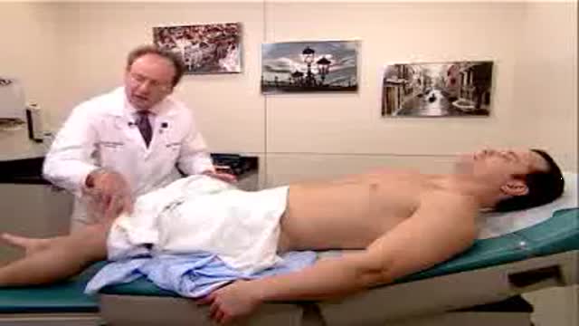
Complete examination of the abdomen including all the items: inspection, palpation, percussion and auscultation Video

Observation of both jugular veins can provide a reliable indication of the volume and pressure in the right side of the heart since internal jugular veins pulsate in response to phasic changes in right atrial pressure. Proper positioning of the patient to increase the effects of gravity enhances distention of the jugular veins and, therefore, increases the ability to observe venous pulsations.

Full complete clinical examination of the chest, lungs and respiration with breath sounds
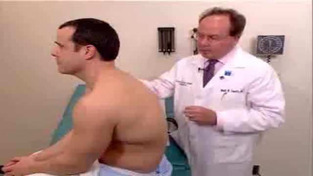
Complete examination of the back

Complete clinical assessment and examination of the neck

Clinical complete examination of the mouth and throat

Complete clinical examination of the ears with all the associated tests

Examination of the eye,vision,retina and field of vision

Examination of the lymph nodes of the head

Optimal blood pressure typically is defined as 120 mm Hg systolic — which is the pressure as your heart beats — over 80 mm Hg diastolic — which is the pressure as your heart relaxes. For your resting heart rate, the target is between 60 and 100 beats per minute (bpm)

The Motor Assessment Scale (MAS) is a performance-based scale that was developed as a means of assessing everyday motor function in patients with stroke (Carr, Shepherd, Nordholm, & Lynne, 1985). The MAS is based on a task-oriented approach to evaluation that assesses performance of functional tasks rather than isolated patterns of movement

Examination of a patient with post-enucleation socket syndrome.
