- Physical Examination
- Surgical Examination
- Ophthalmology
- Clinical Skills
- Orthopedics
- Surgery Videos
- Laparoscopy
- Pediatrics
- Funny Videos
- Cardiothoracic Surgery
- Nursing Videos
- Plastic Surgery
- Otorhinolaryngology
- Histology and Histopathology
- Neurosurgery
- Dermatology
- Pediatric Surgery
- Urology
- Dentistry
- Oncology and Cancers
- Anatomy Videos
- Health and Fitness
- Radiology
- Anaesthesia
- Physical Therapy
- Pharmacology
- Interventional Radiology
- Cardiology
- Endocrinology
- Gynecology
- Emergency Medicine
- Psychiatry and Psychology
- Childbirth Videos
- General Medical Videos
- Nephrology
- Physiology
- Diet and Food Health
- Diabetes Mellitus
- Neurology
- Women Health
- Osteoporosis
- Gastroenterology
- Pulmonology
- Hematology
- Rheumatology
- Toxicology
- Nuclear Medicine
- Infectious Diseases
- Vascular Disease
- Reproductive Health
- Burns and Wound Healing
- Other
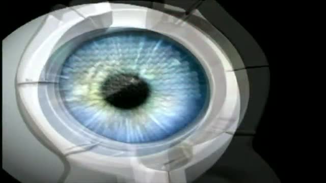
Ophthalmology

Pseudo-exfoliation with small pupil

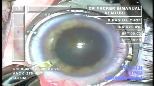
Direct puncture capsulorhexis with a slightly barbed 30g needle on a TB syringe with BSS permits excellent control even with very high vitreous pressure without use of viscoelastic. Ideal for biaxial (microincision) cataract surgery.

Trabeculectomy surgery

Descemet’s stripping automated endothelial keratoplasty (DSAEK) avoids a full-thickness corneal procedure and provides rapid visual rehabilitation. Successful graft positioning while minimizing intraoperative donor endothelial trauma may determine long-term graft survival. Previously described t...echniques for graft insertion may be problematic in some patients with intraoperative floppy iris syndrome (IFIS), anatomically shallow or unstable anterior chambers, or intraoperative increased posterior pressure. This video displays alternative method called the suture drag technique, which may facilitate lamellar endothelial graft insertion under these special circumstances.

To present a new device for fixating the fibro-optic probe during phacoemulsification
Purpose: To evaluate the results of LASIK and IntraLASIK treatment in myopic patients with nystagmus. Methods: Eight patients with congenital nystagmus (16 eyes), from 23 to 49 years of age, underwent LASIK surgery. Corneal flaps were created using either the Hansatome microkeratome or the Intral...ase femtosecond laser. The ablations were performed with the Bausch & Lomb excimer laser with an active tracking system. In some patients, the eyes were fixated with forceps or a fixation ring during the laser ablation. Results: The refractive errors were corrected in all cases. There was no decentration or loss of best corrected visual acuity greater than 1 line. In 56% of the eyes, the post-operative uncorrected visual acuity was better than the best spectacle corrected-visual acuity (BSCVA). 62.5% of the eyes improved their BSCVA. The overall visual performance was improved in all the patients. One patient that did not not drive before become eligible to get a driver license after the surgery. Conclusions: Selected patients with myopia and congenital nystagmus may benefit from laser refractive surgery. Laser refractive surgery may be safely and accurately performed by using either the Hansatome microkeratome or the Intralase femtosecond laser and an active tracking system with or without mechanical fixation. Certain patients improve their BSCVA post-operatively.
Challenges of cataract surgery in the eye with a history of radial keratotomy include IOL power calculation, protection of the cornea and aviodance of capsular complications.

Scleral fixated IOLs in case of inadequacy of capsular support and scleral sutured capsular tension rings when adequate zonular support is inavailable have been recently used in cataract surgery. In these techniques, polypropylene suture is used and the suture ends over the sclera after the knot ha...s been formed, may erode the conjunctiva and become exposed. Thus, the erosion may lead to the development of endophtalmitis. In order to prevent the aforementioned complication, scleral flaps, otologous cornea, duramater or fascia lata patches have been used to cover the knot and rotation of the knot into the tissues has been described.

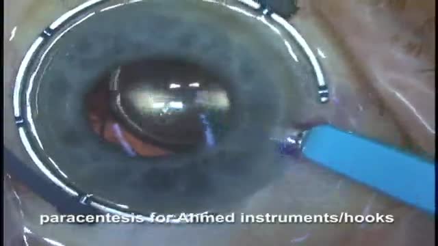
Here we show the placement and removal of the maluygin ring in cataract sugery on a patient on Flomax to control intraoperative floppy iris syndrome (IFIS). The video shows the technique of placing the initial eyelet onto the iris, then toeing down on the inserter to open the lateral eyelets to all...ow them to grad the iris. the trailing haptic is placed with a kuglen hook.

A videos of cataract surgery
Here Drs Oetting and Shriver of the University of Iowa demonstrate the McCannel technique of fixing an IOL to the iris. In this video both the standard McCannel suture retrieval technique and the Siepser/Chang modifed technique are demonstrated. A 10-O prolene with a long curved ctc-6 needle is u...sed to place a suture through the iris and under an 3 piece IOL haptic. Using the standard technique the two ends of the suture are retrieved through a common paracentesis near the fixation site and tied externally. The other haptic is tied using the Siepser sliding knot technique as described by Chang for this indication with an internal knot. The standard technique is a bit easier but does not allow as thight a knot for fixation of the iris to the haptic.

We will present technique of lifting a corneal flap, 10 months post IntraLASIK surgery, after epithelial nest. The nest changed in size and started to grow. The technique is minimal invasive and included partial flap lifting.

The challenge of position a patient with severe kyphosis for cataract extraction and lens implantation is met with a team effort and ingenuity.
Scleral Buckling: Slinging Muscles & Marking Breaks VR1 Basic Techniques
With an Ophthalmoscope, light is shone into the eye and the retina and the optic nerve is examined. This is called as Examination of the Fundus. This is what the eye-doctor sees when he peeps into your eye! Through the transparent cornea, into the dark interior. The Fundus Exam When he looks into the eye with the Ophthalmoscope, he sees a orange glowing interior. That is the retina. The retina is actually transparent. It appears bright because of blood vessels in the choroid layer below. It is like looking at your ear against the bright sunlight. The yellow circle is the Optic Nerve, the cable of vision! A red, shiny dot attracts attention. That is the macula. If indicated, the exam of periphery of the retina is done with an Indirect ophthalmoscope. The ophthalmologist wears this instrument on the head and focuses the light into the eye with a lens held in his hand. This is usually done in a dark room.
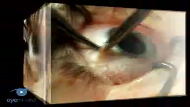
Surgical removal of a Chalazion from the eye lid
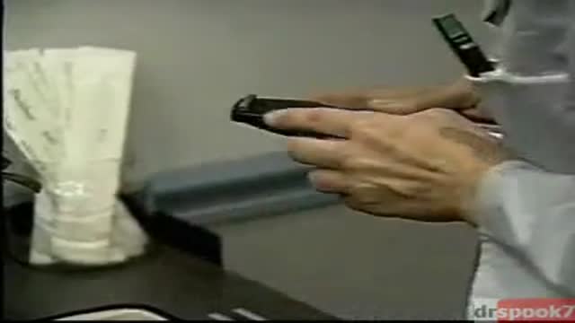
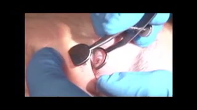
PARASITE REMOVED FROM THE EYEBALL OF A YOUNG N, NOT FOR THE SCREAMISH!Loa Loa worms (also known as the "eye worm") are classified as filarial worms, meaning they thrive in human tissue. The Loa Loa worm is also called the "eye worm" because they often migrate through the eye and surrounding subsurface areas. At one time, prior to the 1920s, loa loa worm infections occurred in the United States. Today, however, they mainly infect people who are native to Sudan, and those who live in or near Central and West Africa's swamps and rain forests.
Loiasis is the infestation of loa loa worms in humans. The larvae are first collected from an infected individual when a mango fly (horsefly) or a deer fly bites the individual, and acquires the larvae. The larvae then progress through the fly's body, finally reaching the feeding tube. They are then transferred to a human host when the fly bites the human. The larvae may remain unnoticed for months or years before becoming an adult, mating, and producing offspring.
Adult female Loa Loa worms can reach a length of 2 1/2 inches while males are approximately half that size. Loa Loa worms can live approximately fifteen years inside their human hosts. They travel continuously through connective and deep tissue, often without the victim experiencing any sensation other than occasional itching.
It is when the worm slows or reaches a sensitive spot that a person will often feel the greatest discomfort. At this point, immune reactions may also include localized redness and a condition called "Calabar" swelling. Skin eruptions and muscle pain may be evident.
When the Loa Loa worm reaches the eye tissue, it can be easily seen and felt within the eyeball for up to an hour. It is usually removed under local anesthesia if the patient is within proximity of a qualified physician. When an adult worm dies, the surrounding tissue may abscess and require excision. Encephalitis can occur if the worm reaches the brain.
After mating, the female will deposit eggs - called microfilariae. These tiny organisms then travel in a worm-like fashion in the bloodstream during daytime hours, when potential host flies are most abundant. They congregate in the lungs at night.
A Loa Loa worm infection is rarely fatal and treatments often cause more life-threatening side effects than the actual infestation, especially if the worms are widespread. The most common treatments are DEC (diethylcarbamazine) and Ivermectin
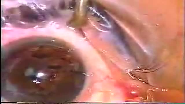
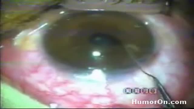
A video of a surgery of corneal graft transplantation
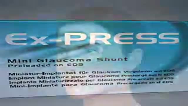
The Ex-PRESS Mini Glaucoma Shunt provides a simplified method of filtration surgery for patients with open angle glaucoma. The Ex-PRESS implanted Under a Scleral Flap is a minimally invasive procedure with predictable results.
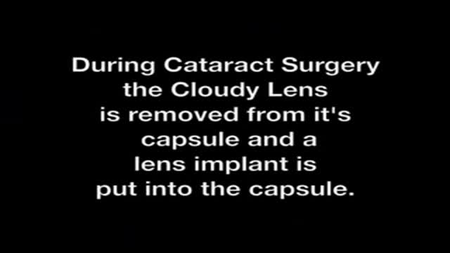
Short Version of Yag Laser Treatment of Capsule Opacity or "after cataract" Video Presentation by Tampa Bay Area Ophthalmologist Ahad Mahootchi, MD from the Eye Clinic of Florida.
