- Physical Examination
- Surgical Examination
- Ophthalmology
- Clinical Skills
- Orthopedics
- Surgery Videos
- Laparoscopy
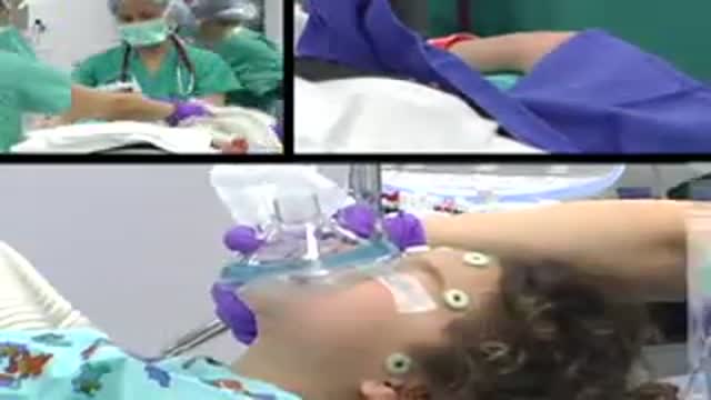
- Pediatrics
- Funny Videos
- Cardiothoracic Surgery
- Nursing Videos
- Plastic Surgery
- Otorhinolaryngology
- Histology and Histopathology
- Neurosurgery
- Dermatology
- Pediatric Surgery
- Urology
- Dentistry
- Oncology and Cancers
- Anatomy Videos
- Health and Fitness
- Radiology
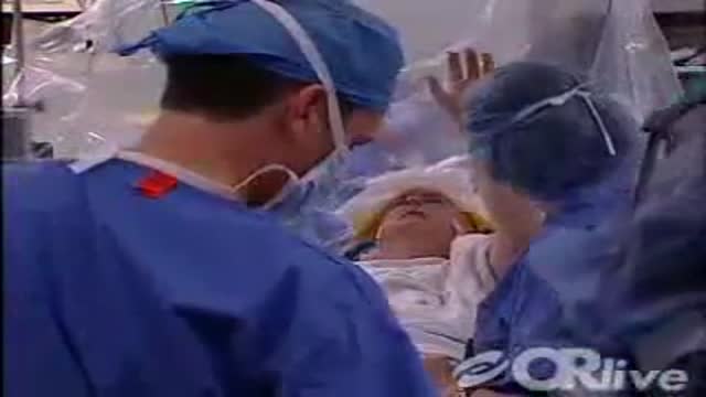
- Anaesthesia
- Physical Therapy
- Pharmacology
- Interventional Radiology
- Cardiology
- Endocrinology
- Gynecology
- Emergency Medicine
- Psychiatry and Psychology
- Childbirth Videos
- General Medical Videos
- Nephrology
- Physiology
- Diet and Food Health
- Diabetes Mellitus
- Neurology
- Women Health
- Osteoporosis
- Gastroenterology
- Pulmonology
- Hematology
- Rheumatology
- Toxicology
- Nuclear Medicine
- Infectious Diseases
- Vascular Disease
- Reproductive Health
- Burns and Wound Healing
- Other
Other

I call this technique deep rendering. I basically stacked graphical cross-sections (in this case, MRI rendering data), using proper increments and clip through them with the camera. This way I am able to explore all internal components in full 3D real-time.
I actually was able to figure out how to colorize different organs to help distinguish them apart from each other but couldn't get the shader to render real-time in Maya.
Credit: MRI scans courtesy of University of Washington Digital Anatomist Program
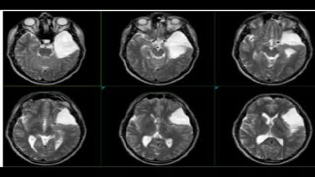
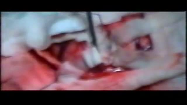
Endoscopic fenestration of suprasellar cyst in a 4 years old girl
Endoscopic fenestration of arachnoid cyst in middle fossa
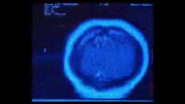
brain scans with arachnoid cyst, pre and post operative

Endoscopic Management of Brain Cyst, ForaminoPlasty

Endoscopic Brain Surgery, third Ventriculostomy
Hydatid Cyst Removal from the brain
Watch as Dr. Benjamin Carson performs risky brain surgery on young Payton to remove a brain tumor. Dr. Carson, director of pediatric neurosurgery, is just one of the many reasons why Johns Hopkins Children's Center was recently ranked #1 in neurology and neurosurgery in America's Best Children's Hospitals 2008
Vanderbilt Medical Center neurosurgeons and neurologists will be online demonstrating their 4-stage innovative technique used for Deep Brain Stimulation (DBS). Deep brain stimulation therapy utilizes an implantable neuro-stimulator to treat movement disorders such as Parkinson's disease, essential tremor, and dystonia.
On Tuesday May 29th at 3:00pm EDT, University Hospitals Case Medical Center Cleveland, Ohio, will host a live webcast to demonstrate the removal of brain tumor and epileptic focus from an awake patient using intra-operative MRI and brain mapping. See this on OR-Live.com
The patient was a middle-aged gentleman with new onset seizures. An MRI showed what appeared to be a low grade glioma near the motor strip on the right. Studies have shown that complete removal can cure the seizures, improve quality of life and survival, but this is difficult to do with conventional technology without harming the surrounding normal brain because its difficult to determine where tumor ends and normal brain begins.

olusegun adekanye's spinal disc replacement operation performed by Dr. Nick Thomas at the Blackheath Hospital.Part 2

olusegun adekanye's spinal disc replacement operation performed by Dr. Nick Thomas at the Blackheath Hospital.
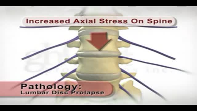
This patient education animation illustrates the internal anatomy of a prolapsed and herniated disc.
A dermatologist explains how this skin condition is recognised and treated and the challenging effects it can have on an individual.
Hair removal techniques have come a long way since the days of messy creams, electrolysis, and shaving. At South Coast MedSpa, we use the most advanced laser technology to do the job efficiently, cleanly, and with minimal discomfort. The SCMS system is fast, gentle, safe, and effective for all skin types and colors.
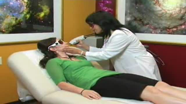
Utilizing specially engineered lasers, permanent hair removal has never been more comfortable for men and women of all colors and skin types. In just four or five sessions, patients can achieve lasting results without damaging the skin or any surrounding tissue.
Dark pigment (melanin) in the hair shaft and the papilla (the root of the hair follicle) are targeted by a specific light-energy emitted by the laser. In a tiny fraction of a second, the hair is simply vaporized without damaging the skin or any surrounding tissue.
In one pulse (that lasts a tiny fraction of a second) our lasers remove hair on a patch of skin the size of a quarter. The hair removal sensation is like plucking hair or getting snapped by a rubber-band. Our lasers incorporate a patented and state-of-the-art integrated cooling system that acts as a natural anesthetic, cooling down the skin to minimize any discomfort. Patients unanimously report that the hair removal treatment is a "piece of cake" compared to waxing.
Cerebellar functions of the lower limbs from the USMLE collection
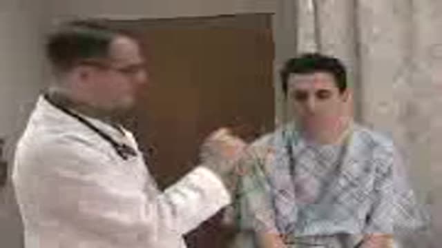
Cerebellar functions of the upper limbs from the USMLE collection
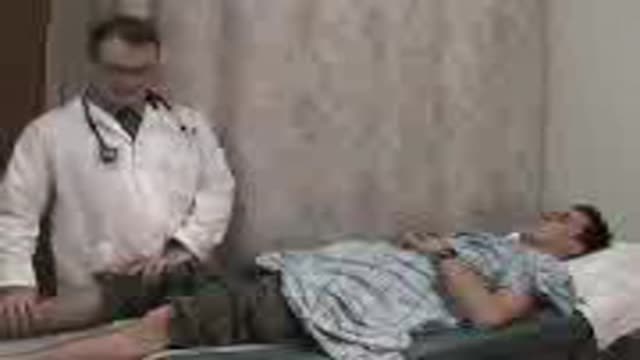
Meningeal Irritation Signs from the USMLE collection
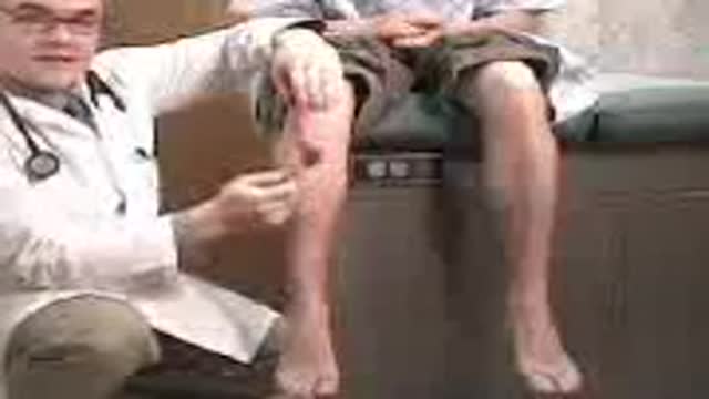
Knee reflex video from the USMLE collection
