- Physical Examination
- Surgical Examination
- Ophthalmology
- Clinical Skills
- Orthopedics
- Surgery Videos
- Laparoscopy
- Pediatrics
- Funny Videos
- Cardiothoracic Surgery
- Nursing Videos
- Plastic Surgery
- Otorhinolaryngology
- Histology and Histopathology
- Neurosurgery
- Dermatology
- Pediatric Surgery
- Urology
- Dentistry
- Oncology and Cancers
- Anatomy Videos
- Health and Fitness
- Radiology
- Anaesthesia
- Physical Therapy
- Pharmacology
- Interventional Radiology
- Cardiology
- Endocrinology
- Gynecology
- Emergency Medicine
- Psychiatry and Psychology
- Childbirth Videos
- General Medical Videos
- Nephrology
- Physiology
- Diet and Food Health
- Diabetes Mellitus
- Neurology
- Women Health
- Osteoporosis
- Gastroenterology
- Pulmonology
- Hematology
- Rheumatology
- Toxicology
- Nuclear Medicine
- Infectious Diseases
- Vascular Disease
- Reproductive Health
- Burns and Wound Healing
- Other
Latest videos

Mysterious things happen in nature, and extraordinary birth delivery facts amaze and astound us. And "The baby who didn't know he was born" is one of them; the reason was because his mother didn't break water, so the little one thought was still in the womb. Of course, the amniotic sac was later broken by the doctor, and as soon as this happened the baby began to breath and cry.

10 Biggest Babies Ever Born
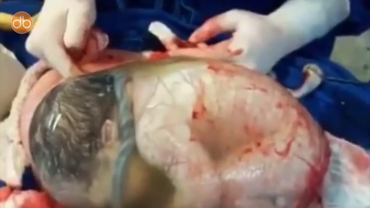
Baby Born Still Inside The Amniotic Sac

Cerebral hydatid disease (neurohydatidosis) is caused by Echinococcus granulosus or less commonly E. alveolaris or E. multilocularis. The larval stage is the cause of hydatid disease in humans 1. Epidemiology Cerebral hydatid disease is a rare parasitic infestation and accounts for 1-2 % of all cystic echinococcosis. Hydatid disease is endemic in the Mediterranean region, the Middle East, Africa, eastern part of Turkey, Australia and parts of South America 2. Clinical presentation Symptoms and signs include: focal neurological deficits headaches increased intracranial pressure hydrocephalus papilloedema and loss of vision altered mental status seizures (rare)

Most of us have taken a sex education class or two. We know what condoms are supposed to be used for. Whether or not people use condoms every time they are necessary is a totally different story. You were probably taught the necessary but embarrassing lesson of how to put a condom on by the visual aids your sex education teacher provided. Of course, these tactics are a little more modern, so depending on how old you are, you may not have learned the basics of condom use until after high school. Yes, condoms are a pretty smart invention and they’re pretty safe to use. They are over 90% effective against sexually transmitted diseases (STDs), and of course, they help to reduce the risk of pregnancy by 98%. Some guys claim that they’re not comfortable to wear, which is why some companies have come up with new condoms that have a more natural fit and provide pleasure for both partners. However, with all the things we know about condoms, there’s still so much we don’t know. Here are 10 facts about condoms that are just as interesting as the condoms themselves.
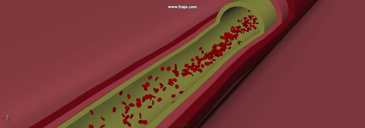
The heart and circulatory system (also called the cardiovascular system) make up the network that delivers blood to the body's tissues. With each heartbeat, blood is sent throughout our bodies, carrying oxygen and nutrients to all of our cells.
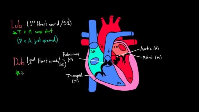
The cardiac conduction system is a group of specialized cardiac muscle cells in the walls of the heart that send signals to the heart muscle causing it to contract. The main components of the cardiac conduction system are the SA node, AV node, bundle of His, bundle branches, and Purkinje fibers.
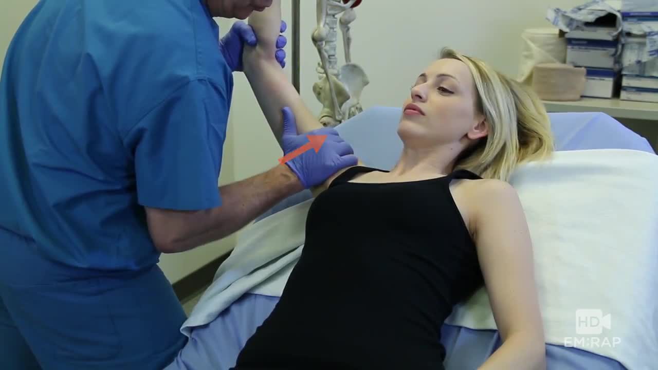
A technique for reducing an inferior shoulder dislocation. watch to learn more
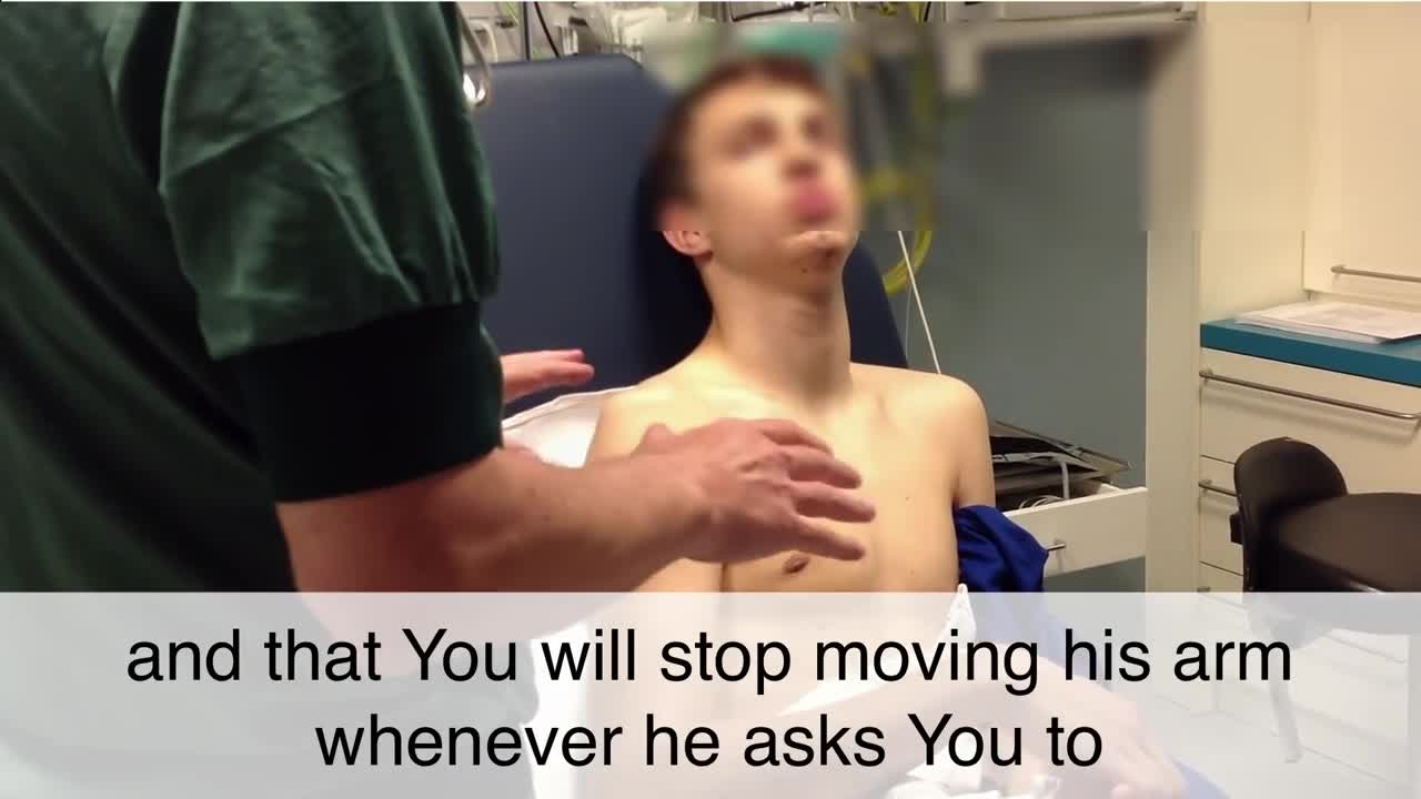
Reduction techniques can vary in terms of required force, time, equipment, and staff. [7] No single reduction method is successful in every instance; therefore, the clinician should be familiar with several reduction techniques. Techniques commonly used to reduce anterior shoulder dislocations include the following [35, 36, 37, 38, 39] : Stimson maneuver Scapular manipulation External rotation Milch technique Spaso technique Traction-countertraction
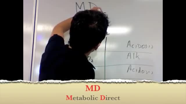
Here's a quick simple way to determine if a pH disturbance is respiratory or metabolic.
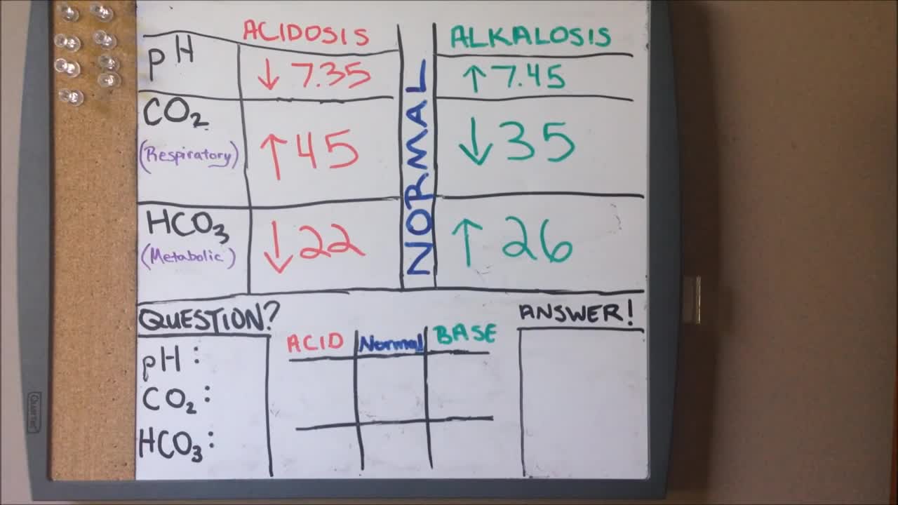
ABGs Made Easy | Arterial Blood Gas | Acid Base Balance: Everything You Need To Know!
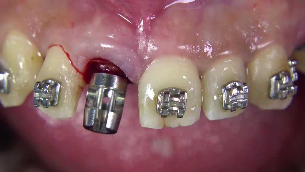
Benex II Surgical Extraction System
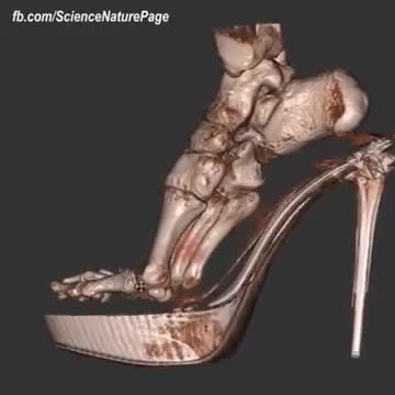
What happens when you wear High Heels. SHOW MORE

The pain is your feet trying to tell you something!
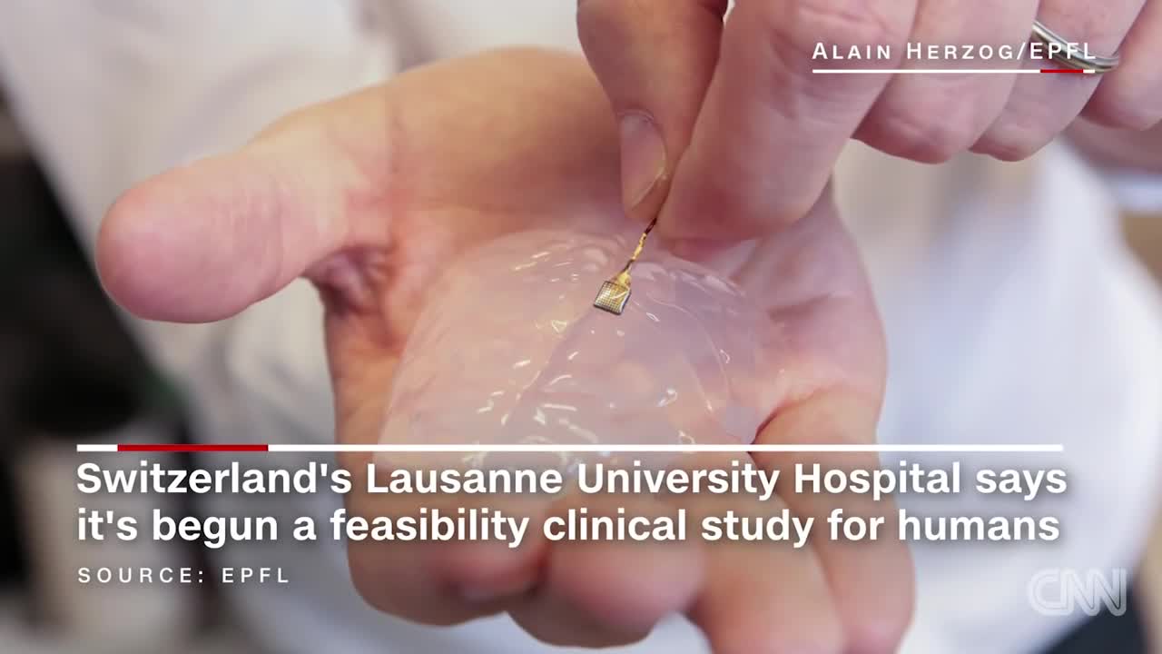
Scientists have developed a wireless brain implant that enabled a paralyzed monkey to walk again.
Coronary artery vasospasm, or smooth muscle constriction of the coronary artery, is an important cause of chest pain syndromes that can lead to myocardial infarction (MI), ventricular arrhythmias, and sudden death. It also plays a key role in the development of atherosclerotic lesions.Nov 22, 2016
Prinzmetal's or Prinzmetal angina (/ˈprɪntsmɛtəl/, sounds like "prints metal") (also known as variant angina, vasospastic angina (VSA), angina inversa, or coronary vessel spasm) is a syndrome typically consisting of angina (cardiac chest pain) at rest that occurs in cycles.
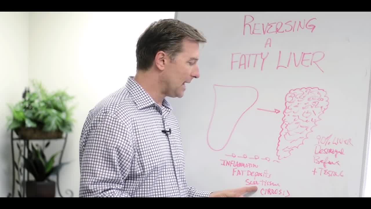
explains about fatty liver symptoms and fatty liver treatment. watch to learn more

Fatty liver is a dangerous yet misunderstood disease. In America, it affects 90 million of us and 17 percent of our children.
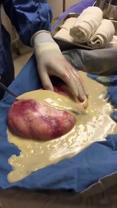
Ovarian teratoma is a type of germ cell tumour. Germ cell tumours are cancers that begin in egg cells in women or sperm cells in men. There are 2 main types of ovarian teratoma. Mature teratoma, which is benign. Immature teratoma, which is cancerous.
