- Physical Examination
- Surgical Examination
- Ophthalmology
- Clinical Skills
- Orthopedics
- Surgery Videos
- Laparoscopy
- Pediatrics
- Funny Videos
- Cardiothoracic Surgery
- Nursing Videos
- Plastic Surgery
- Otorhinolaryngology
- Histology and Histopathology
- Neurosurgery
- Dermatology
- Pediatric Surgery
- Urology
- Dentistry
- Oncology and Cancers
- Anatomy Videos
- Health and Fitness
- Radiology
- Anaesthesia
- Physical Therapy
- Pharmacology
- Interventional Radiology
- Cardiology
- Endocrinology
- Gynecology
- Emergency Medicine
- Psychiatry and Psychology
- Childbirth Videos
- General Medical Videos
- Nephrology
- Physiology
- Diet and Food Health
- Diabetes Mellitus
- Neurology
- Women Health
- Osteoporosis
- Gastroenterology
- Pulmonology
- Hematology
- Rheumatology
- Toxicology
- Nuclear Medicine
- Infectious Diseases
- Vascular Disease
- Reproductive Health
- Burns and Wound Healing
- Other
Latest videos
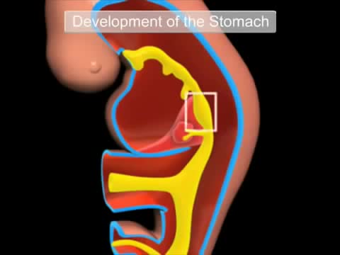
The gastrointestinal tract (GIT) arises initially during the process of gastrulation from the endoderm of the trilaminar embryo (week 3) and extends from the buccopharyngeal membrane to the cloacal membrane. The tract and associated organs later have contributions from all the germ cell layers. During the 4th week three distinct regions (fore-, mid- and hind-gut) extend the length of the embryo and will contribute different components of the GIT. The large mid-gut is generated by lateral embryonic folding which "pinches off" a pocket of the yolk sac, the 2 compartments continue to communicate through the vitelline duct. The oral cavity (mouth) is formed following breakdown of the buccopharyngeal membrane (oropharyngeal or oral membrane) and contributed to mainly by the pharynx lying within the pharyngeal arches (More? Head Development). Loss of buccopharyngeal membrane opens the tract to amniotic fluid through the remainder of development, and during the fetal period is actively swallowed.

Gastroschisis is a birth defect that develops in a baby while a woman is pregnant. This condition occurs when an opening forms in the baby's abdominal wall. The baby's bowel pushes through this hole. It then develops outside of the baby's body in the amniotic fluid.
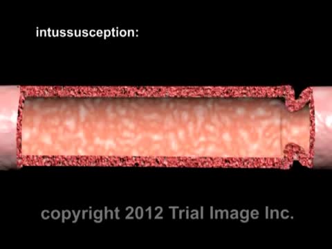
Intussusception (in-tuh-suh-SEP-shun) is a serious condition in which part of the intestine slides into an adjacent part of the intestine. This "telescoping" often blocks food or fluid from passing through. Intussusception also cuts off the blood supply to the part of the intestine that's affected, which can lead to a tear in the bowel (perforation), infection and death of bowel tissue.
An omphalocele is a birth defect in which an infant's intestine or other abdominal organs are outside of the body because of a hole in the belly button (navel) area. The intestines are covered only by a thin layer of tissue and can be easily seen.
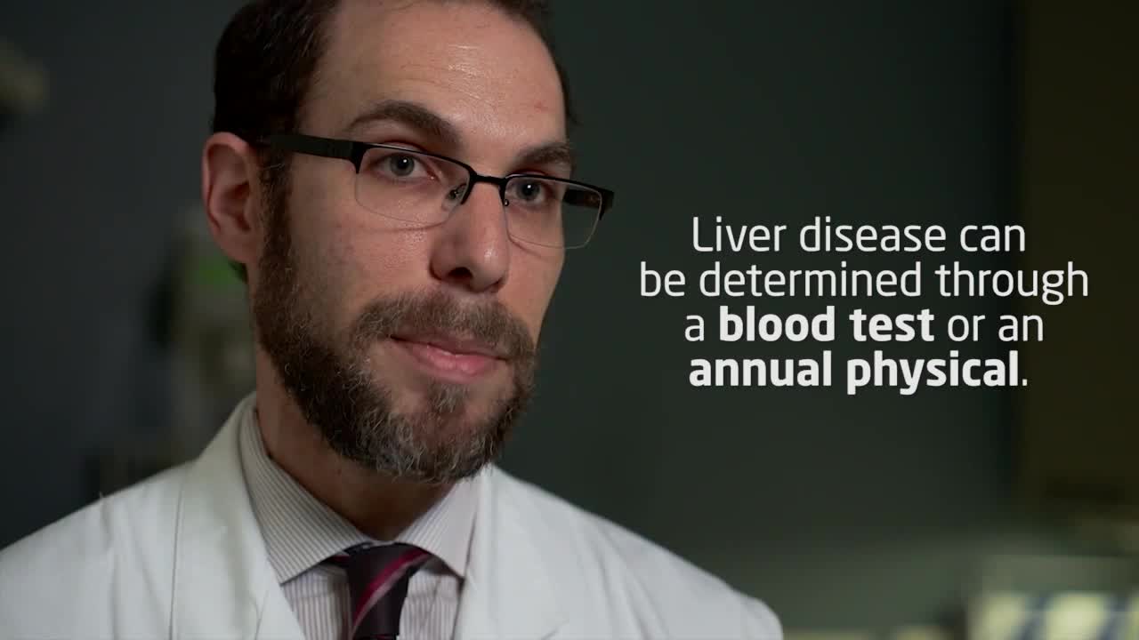
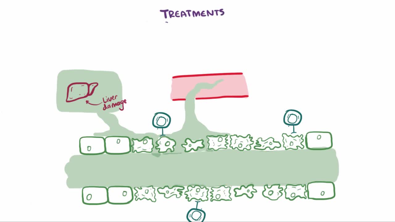
Alcoholic liver disease is a term that encompasses the liver manifestations of alcohol overconsumption, including fatty liver, alcoholic hepatitis, and chronic hepatitis with liver fibrosis or cirrhosis. It is the major cause of liver disease in Western countries.
As the liver becomes more severely damaged, more obvious and serious symptoms can develop, such as: yellowing of the skin and whites of the eyes (jaundice) swelling in the legs, ankles and feet, due to a build-up of fluid (oedema) swelling in your abdomen, due to a build-up of fluid known as ascites.
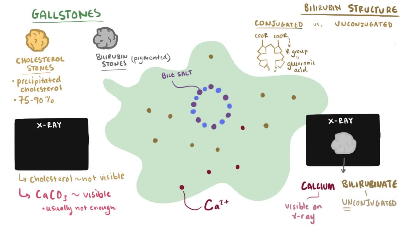
Cholelithiasis involves the presence of gallstones (see the image below), which are concretions that form in the biliary tract, usually in the gallbladder. Choledocholithiasis refers to the presence of 1 or more gallstones in the common bile duct (CBD).
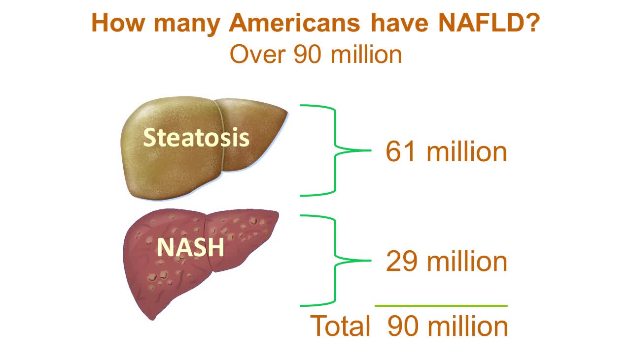
Nonalcoholic fatty liver disease is an umbrella term for a range of liver conditions affecting people who drink little to no alcohol. As the name implies, the main characteristic of nonalcoholic fatty liver disease is too much fat stored in liver cells. Nonalcoholic steatohepatitis, a potentially serious form of the disease, is marked by liver inflammation, which may progress to scarring and irreversible damage. This damage is similar to the damage caused by heavy alcohol use. At its most severe, nonalcoholic steatohepatitis can progress to cirrhosis and liver failure Nonalcoholic fatty liver disease is increasingly common around the world, especially in Western nations. In the United States, it is the most common form of chronic liver disease, affecting an estimated 80 to 100 million people. Nonalcoholic fatty liver disease occurs in every age group but especially in people in their 40s and 50s who are at high risk of heart disease because of such risk factors as obesity and type 2 diabetes. The condition is also closely linked to metabolic syndrome, which is a cluster of abnormalities including increased abdominal fat, poor ability to use the hormone insulin, high blood pressure and high blood levels of triglycerides, a type of fat. Nonalcoholic fatty liver disease care at Mayo Clinic Request an Appointment at Mayo Clinic Symptoms & causes Aug. 23, 2016 Print Share on: Facebook Twitter References Related Magnetic resonance elastography Nonalcoholic fatty liver disease Overview Symptoms & causes Diagnosis & treatment Diagnosis Treatment Departments & specialties Expertise & rankings Locations, travel & lodging Clinical trials Research Costs & insurance Preparing for your appointment Self-management More about In-Depth Multimedia Resources News from Mayo Clinic Advertisement

Gitelman syndrome is a kidney disorder that causes an imbalance of charged atoms (ions) in the body, including ions of potassium, magnesium, and calcium. The signs and symptoms of Gitelman syndrome usually appear in late childhood or adolescence. Common features of this condition include painful muscle spasms (tetany), muscle weakness or cramping, dizziness, and salt craving. Also common is a tingling or prickly sensation in the skin (paresthesias), most often affecting the face. Some individuals with Gitelman syndrome experience excessive tiredness (fatigue), low blood pressure, and a painful joint condition called chondrocalcinosis. Studies suggest that Gitelman syndrome may also increase the risk of a potentially dangerous abnormal heart rhythm called ventricular arrhythmia. The signs and symptoms of Gitelman syndrome vary widely, even among affected members of the same family. Most people with this condition have relatively mild symptoms, although affected individuals with severe muscle cramping, paralysis, and slow growth have been reported.
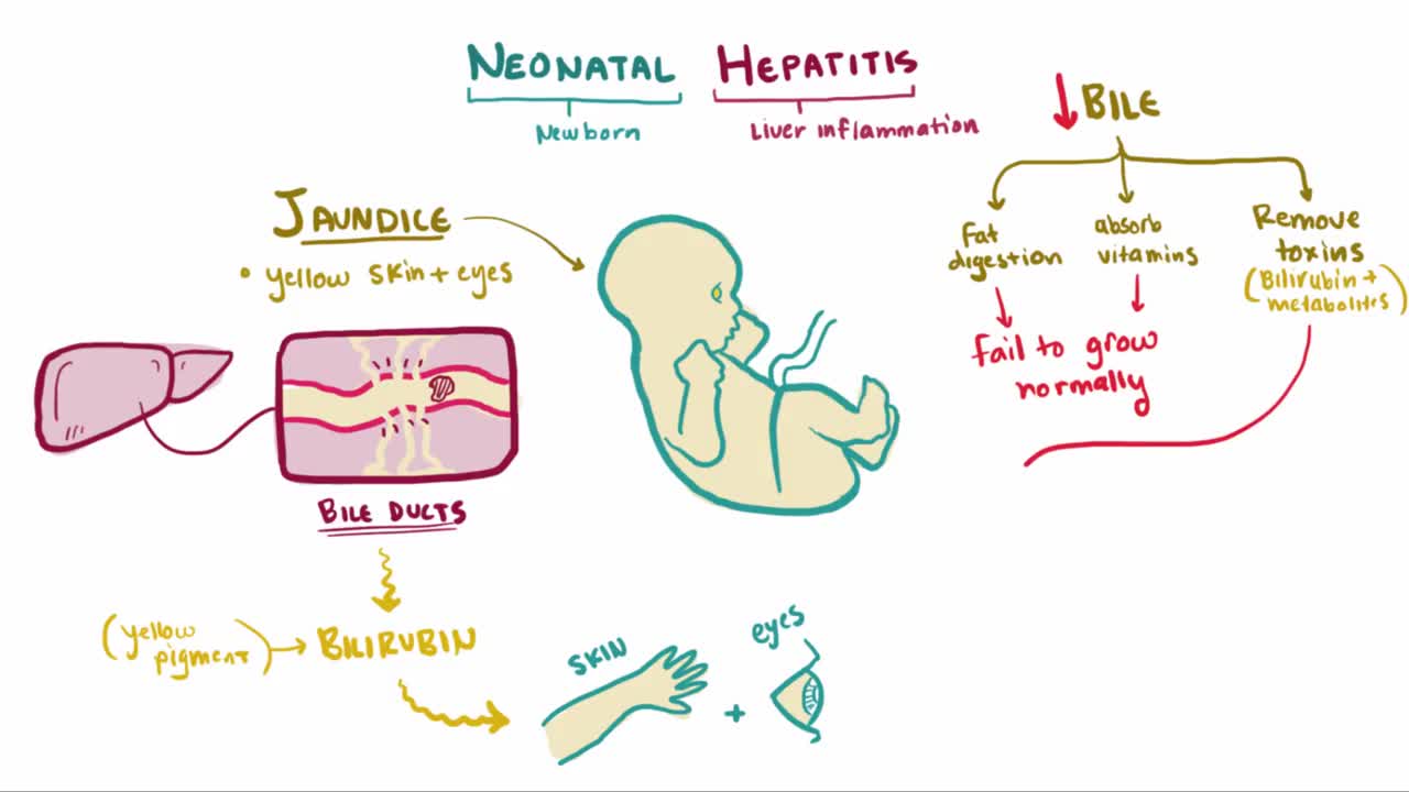
What is neonatal hepatitis? Neonatal hepatitis is an inflammation of an infant's liver just after birth, sometimes this inflammation is due to a virus but in most cases the cause is unknown, or idiopathic
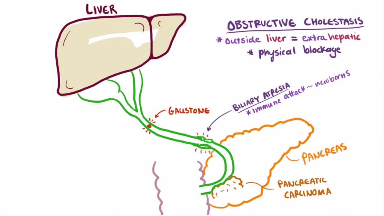
Cholestatic liver disease is a condition that results from an impairment of bile formation or bile flow to the gallbladder and duodenum (first section of the small intestine). ... The effects of cholestasis are profound and widespread, leading to worsening liver disease and systemic illness.
Primary biliary cholangitis (PBC), formerly known as primary biliary cirrhosis, is a chronic liver disease resulting from progressive destruction of the bile ducts in the liver – called the intrahepatic bile ducts. Bile produced in your liver travels via these ducts to your small intestine where it aids in the digestion of fat and fat-soluble vitamins (A, D, E and K). When the ducts are destroyed, bile builds up in the liver contributing to inflammation and scarring (fibrosis). Eventually this can lead to cirrhosis and its associated complications, as scar tissue replaces healthy liver tissue and liver function becomes increasingly impaired.
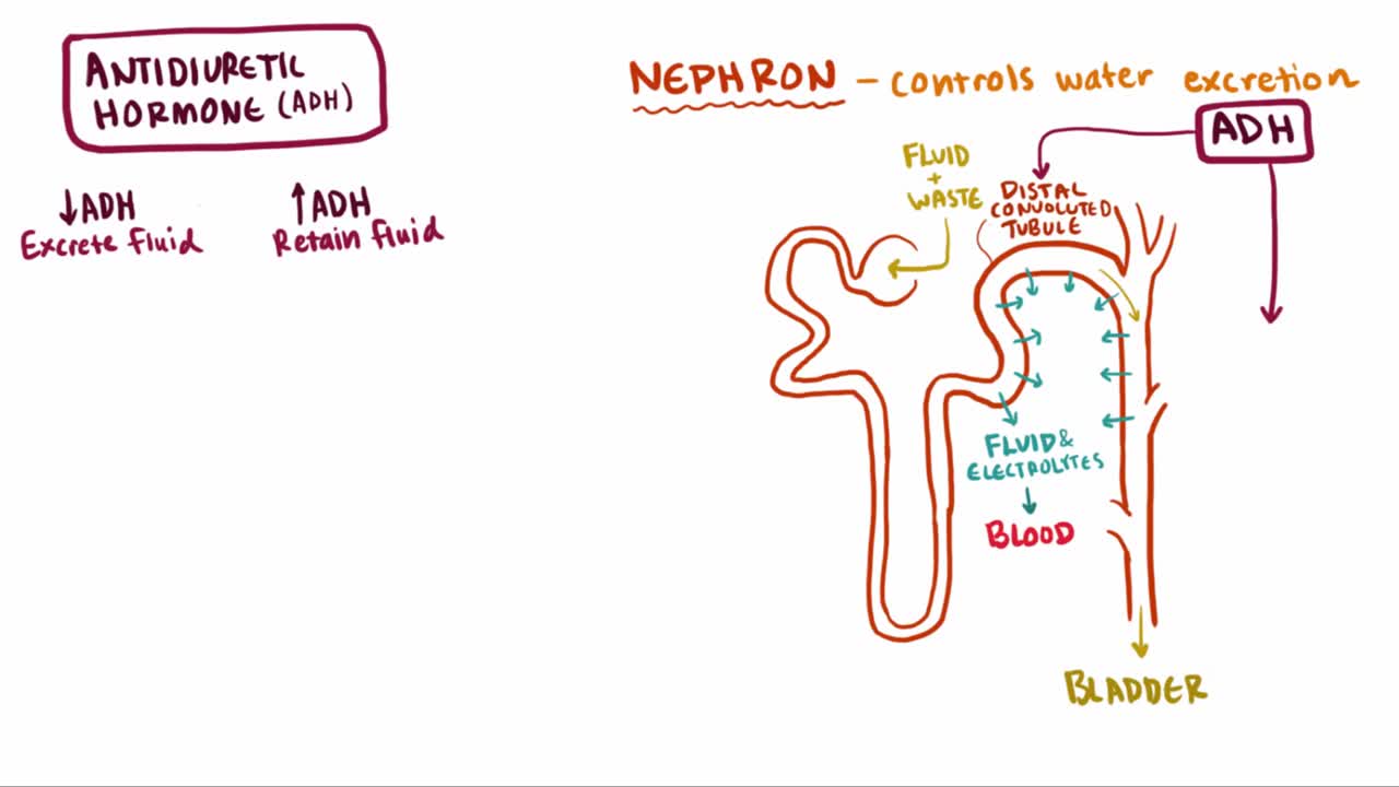
What is syndrome of inappropriate antidiuretic hormone (SIADH)? Well, SIADH is a condition where too much ADH hormone is released, which causes an increase in blood volume and ultimately leads to a series of complications related to the blood osmolality and osmolarity
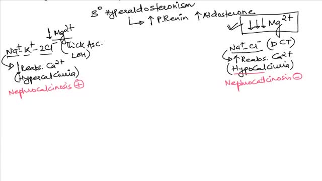
Bartter syndrome has traditionally been classified into three main clinical variants, as follows: Neonatal (or antenatal) Bartter syndrome Classic Bartter syndrome Gitelman syndrome Advances in molecular diagnostics have revealed that Bartter syndrome results from mutations in numerous genes that affect the function of ion channels and transporters that normally mediate transepithelial salt reabsorption in the distal nephron segments. Hundreds of mutations have been identified to date. Such advances may result in the development of new therapies (see the image below). [2] (See Pathophysiology and Etiology.)
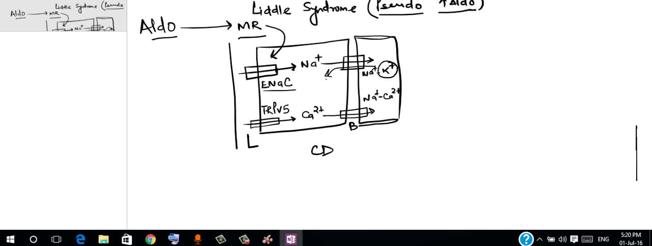
Liddle syndrome is an inherited form of high blood pressure (hypertension). This condition is characterized by severe hypertension that begins unusually early in life, often in childhood, although some affected individuals are not diagnosed until adulthood. Some people with Liddle syndrome have no additional signs or symptoms, especially in childhood. Over time, however, untreated hypertension can lead to heart disease or stroke, which may be fatal.
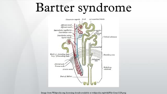
Bartter syndrome, originally described by Bartter and colleagues in 1962, [1] represents a set of closely related, autosomal recessive renal tubular disorders characterized by hypokalemia, hypochloremia, metabolic alkalosis, and hyperreninemia with normal blood pressure. The underlying renal abnormality results in excessive urinary losses of sodium, chloride, and potassium.
Formerly called toxemia, preeclampsia is a condition that pregnant women develop. It is marked by high blood pressure in women who have previously not experienced high blood pressure before. Preeclamptic women will have a high level of protein in their urine and often also have swelling in the feet, legs, and hands. This condition usually appears late in pregnancy, generally after the 20 week mark, although it can occur earlier

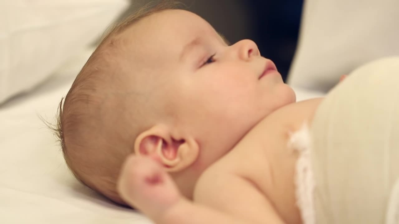
Developmental Psychology Documentary on Brain and Intelligence Development in Babies SHOW MORE

Scientists have found that every baby has genius potential, a child's education must begin early in order to develop the potential it has. Pregnancy is not too early to start, as evidence indicating that the developing fetus can learn is ever mounting.

uses video of babies and toddlers to show the communication milestones expected in typically developing children. She also discusses what parents should do if they suspect their child is developmentally delayed
