- Physical Examination
- Surgical Examination
- Ophthalmology
- Clinical Skills
- Orthopedics
- Surgery Videos
- Laparoscopy
- Pediatrics
- Funny Videos
- Cardiothoracic Surgery
- Nursing Videos
- Plastic Surgery
- Otorhinolaryngology
- Histology and Histopathology
- Neurosurgery
- Dermatology
- Pediatric Surgery
- Urology
- Dentistry
- Oncology and Cancers
- Anatomy Videos
- Health and Fitness
- Radiology
- Anaesthesia
- Physical Therapy
- Pharmacology
- Interventional Radiology
- Cardiology
- Endocrinology
- Gynecology
- Emergency Medicine
- Psychiatry and Psychology
- Childbirth Videos
- General Medical Videos
- Nephrology
- Physiology
- Diet and Food Health
- Diabetes Mellitus
- Neurology
- Women Health
- Osteoporosis
- Gastroenterology
- Pulmonology
- Hematology
- Rheumatology
- Toxicology
- Nuclear Medicine
- Infectious Diseases
- Vascular Disease
- Reproductive Health
- Burns and Wound Healing
- Other
Latest videos
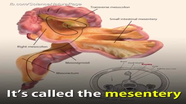
There is a new organ in your digestive system SHOW MORE
A Chinese hospital in the process of creating a human ear almost entirely through the human anatomy alone.
Craft man’s new ear from rib cartilage and the skin on his forearm

knife spoon and toothbrush removed from stomach
Super Obese individuals (people with a Body Mass Index over 45) have an increased risk during any surgery. And the longer the time under anesthesia, the greater the risk. Gastric bypass surgery can last over 2 hours. Duodenal switch surgery often takes over 4 hours. That’s a long time to be under anesthesia.
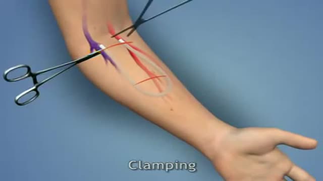
A surgeon creates an arteriovenous fistula by making a connection between an artery (which carries blood away from the heart) and a vein (which carries blood back to the heart). This artificial connection allows the vein to become larger and for the walls of the vein to thicken, a process termed maturation. A mature fistula makes it easier for the vein to be punctured repeatedly for dialysis. Maturation typically takes three to six months to occur, but in rare cases, can take up to a year. This makes advance planning for an arteriovenous fistula important. When a patient is felt to be approximately a year away from requiring dialysis, the patient should be referred for evaluation for possible creation of an arteriovenous fistula.
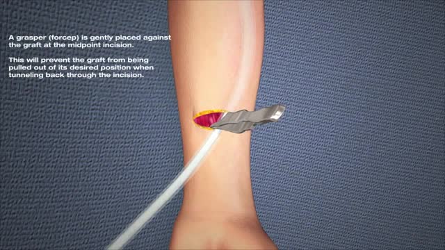
Hemodialysis, also called dialysis, is the most common treatment for kidney failure. A dialysis machine is an artificial kidney which cleanses the blood. During dialysis, blood is drawn from the patient into the dialysis machine, circulated through the machine, and then returned to the patient. Two needles are inserted into the patient's bloodstream to allow this process to occur. Hemodialysis is normally performed three times a week and the purpose of vascular access is to provide reliable sites where the bloodstream can be easily accessed each time. There are three major types of vascular access: arteriovenous fistula, arteriovenous graft, and venous catheter. The great majority of vascular accesses are created in the arm, but they can also be created in the leg.
Your kidneys are two bean-shaped organs that lie just below your rib cage, on each side of your spine. They remove waste from your body, level out your blood pressure, and keep your bones strong. They also ensure that you have the right amount of chemicals, like potassium and sodium (salt), in your blood. Finally, they make the hormone that causes your body to create red blood cells.
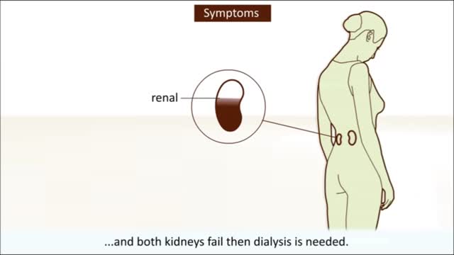
Dialysis and kidney transplantation are treatments for severe kidney failure, also called kidney (or renal) failure, stage 5 chronic kidney disease, and end-stage kidney (or renal) disease. There are two types of dialysis: hemodialysis and peritoneal dialysis. When the kidneys are no longer working effectively, waste products, electrolytes, and fluid build up in the blood. Dialysis takes over a portion of the function of the failing kidneys to remove the fluid and waste products. Kidney transplantation can more completely take over the function of the failing kidneys.

Haemorrhoids is one of the most common problems seen in surgical OPD. Open haemorrhoidectomy has remained the gold standard for a long time with a high post-operative morbidity. The quest for a better understanding of the pathology of haemorrhoids resulted in the evolvement of stapler haemorrhoidopexy. Our aim is to study the efficacy of stapler haemorrhoidopexy with regards to role of immediate post-operative morbidity. A prospective study of 50 patients (n = 50) with the second- and third-degree symptomatic haemorrhoids was done. The mean age of the patients was 44.1 years. Fourteen patients had co-morbid conditions. The average duration of the operation was 29 min. Patients with the second-degree haemorrhoids had higher rate of complication. The complication rate was 32%. Three patients had urinary retention. Two patients had minor bleeding, and one patient experienced transient discharge. The mean analgesic requirement was 2.4 tramadol, 50 mg injections. Ten patients had significant post-operative pain. Average length of hospital stay was 2.7 days. There were no symptomatic recurrences till date.
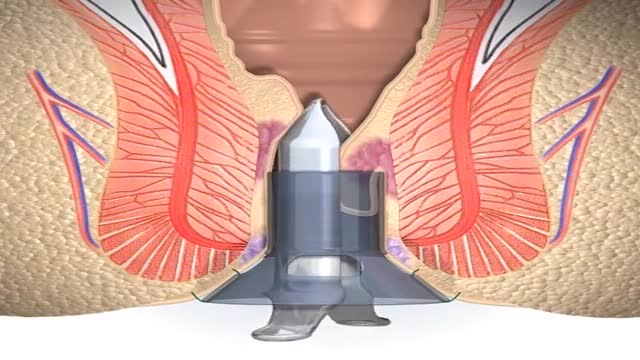
A stapled haemorrhoidopexy is an operation to return the haemorrhoids to a normal. position inside the rectum (back passage). A circular shaped stapling device is gently. inserted in the back passage. The surgeon is then able to use the device to remove.

Twins in the Womb - Human Development

Twins Conversation

Womb Fight amazing

In the Womb - Identical Twins

MRI Shows Twins Fighting in the Womb
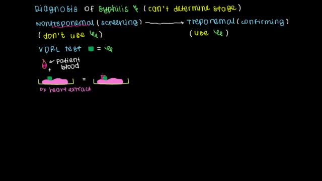
Learn what tests can be used to screen and diagnose syphilis as well as how to treat and prevent the infection.
Syphilis develops in stages, and symptoms vary with each stage. But the stages may overlap, and symptoms don't always occur in the same order. You may be infected with syphilis and not notice any symptoms for years.

Syringomyelia is a cystic cavitation of the spinal cord associated with Chiari I malformation (70%) or basilar invagination (10%) or tumor. It may be a post-traumatic condition. There are 2 main forms: communicating with the central canal or subarachnoid spaces (Chiari I malformation); non communicating (trauma, tumors).
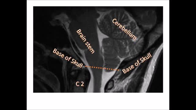
Chiari malformation (kee-AH-ree mal-for-MAY-shun) is a condition in which brain tissue extends into your spinal canal. It occurs when part of your skull is abnormally small or misshapen, pressing on your brain and forcing it downward.
