Neueste Videos

Mesenteric cyst is one of the rarest abdominal tumours, with approximately 820 cases reported since 1507. The incidence varies from 1 per 100,000 to 250,000 admissions. The lack of characteristic clinical features and radiological signs may present great diagnostic difficulties.
Suspect that a patient has a subphrenic abscess if he deteriorates, or recovers and then deteriorates, between the 14th and the 21st day after a laparotomy, with a low, slowly increasing, swinging fever, sweating, and a tachycardia. This, and a leucocytosis, show that he has ''pus somewhere', which is making him anorexic, wasted, and ultimately cachectic. If he has no sign of a wound infection, a rectal examination is negative, and his abdomen is soft and relaxed, the pus is probably under his diaphragm. The pus might be between his diaphragm and his liver, in (1) his right or (2) his left subphrenic space, or under his liver in (3) his right or (4) his left subhepatic space in his lesser sac. He may have pus in more than one of these spaces. Explore him on the suspicion that he might have a subphrenic abscess. Exploration is not a major operation; the difficulty is knowing where to explore, so refer him if you can. If you cannot refer him, explore him yourself. If you fail to find pus, you have done him no harm; missing a subphrenic abscess is far worse. If it is anterior, you can drain it by going under his costal margin anteriorly. If it is posterior, you can go through the bed of his 12th rib posteriorly.

Of the many factors that affect your compatibility with a man, one of the biggest (or smallest) is in his pants. As with humour, interests or habits, the wrong fit can leave you cold. Or traumatised. In a study of 1,661 penises, Dr Debby Herbenick, author of Sex Made Easy, found an almost nine-inch difference in erection size: from 1.6 inches to 10.2. And since absolutely nothing outside the package tells you what to expect with the package, you have to test compatibility the hard way. Sometimes you hit your jackpot, sometimes it's just fine, and sometimes he's the guy on either end of that erection spectrum. These writers have been there, so here's what they learned - and how you can deal (without the gasp reflex).

Do "natural male enhancements" and mail-order sexual function drugs work? No. That's according to Dr. LeRoy Jones a urologist and men's health specialist with Urology San Antonio.

Erectile dysfunction (ED) is the inability to get or keep an erection firm enough for sexual function. It’s a common sexual problem, affecting as many as 30 million men in the United States. Most cases of ED have a physical cause, such as heart disease, diabetes, and obesity. Lifestyle choices like smoking and drinking excessive amounts of alcohol can also lead to ED. But for some men, psychological issues are the root of the problem.

Inducing anesthesia (lack of sensation or feeling) before surgery or certain procedures that do not require skeletal muscle relaxation. It may also be used for other conditions as determined by your doctor.

Phencyclidine (PCP) was developed in the 1950s as an intravenous anesthetic but, due to the side effects of confusion and delirium, its development for human medical use was discontinued. In its pure form, it is a white crystalline powder that readily dissolves in water or alcohol and has a distinctive bitter chemical taste. On the illicit drug market, Phencyclidine contains a number of contaminants as a result of makeshift manufacturing, causing the color to range from tan to brown, and the consistency to range from powder to a gummy mass. It is available in a variety of tablets, capsules, and colored powders, which are either taken orally or snorted. The liquid form of phencyclidine is actually phencyclidine base dissolved most often in ether, a highly flammable solvent. For smoking, phencyclidine is typically sprayed onto leafy material such as mint, parsley, oregano, or marijuana.

LSD is one of the most potent, mood-changing chemicals. It is manufactured from lysergic acid, which is found in the ergot fungus that grows on rye and other grains. It is produced in crystal form in illegal laboratories, mainly in the United States. These crystals are converted to a liquid for distribution. It is odorless, colorless, and has a slightly bitter taste.
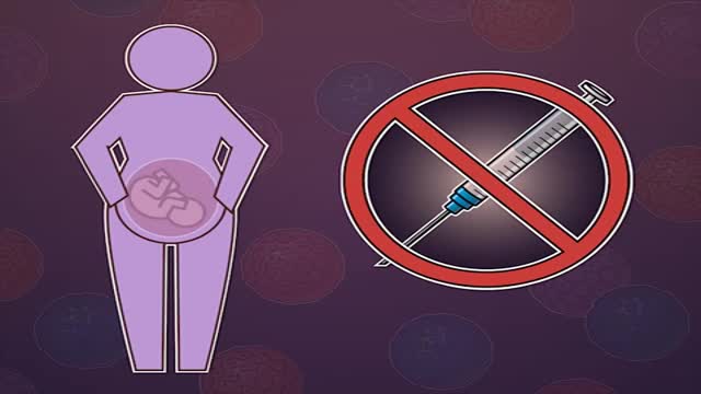
HPV causes genital warts and cervical and other anogenital cancers. The HPV vaccine is recommended for girls and women 9 to 26 years of age to reduce infections, but information on safety in pregnant women is limited.
What happens to our bodies after we die?

Smallpox disease is a serious, highly contagious, and often life-threatening infection marked by a rash of round pox (blisters) on the face, arms, and legs. It is caused by the Variola virus. The last case of smallpox in the United States was in 1949.

Wetness. Even the most absorbent diaper leaves some moisture on your child's skin. And when your child's urine mixes with bacteria from his stool, it breaks down into ammonia, which can be very harsh on the skin. That's why children with frequent bowel movements or diarrhea are more prone to diaper rash.

Eczema, or atopic dermatitis, is a rash that primarily occurs in people with asthma or allergies. The rash is often reddish and itchy with a scaly texture. Psoriasis is a common skin condition that can cause a scaly, itchy, red rash to form along the scalp, elbows, and joints.Apr 13, 2016

Classical PKU is an autosomal recessive disorder, caused by mutations in both alleles of the gene for phenylalanine hydroxylase (PAH), found on chromosome 12. In the body, phenylalanine hydroxylase converts the amino acid phenylalanine to tyrosine, another amino acid.

PKU is inherited in families in an autosomal recessive pattern. Autosomal recessive inheritance means that a person has two copies of the gene that is altered. Usually, each parent of an individual who has PKU carries one copy of the altered gene. ... Gene alterations (mutations) in the PAH gene cause PKU.

If you have gestational diabetes, your baby may be at increased risk of: Excessive birth weight. Extra glucose in your bloodstream crosses the placenta, which triggers your baby's pancreas to make extra insulin. This can cause your baby to grow too large (macrosomia).

Because the continuous supply of glucose is stopped after birth, the neonate develops hypoglycemia because of insufficient substrate. Stimulation of fetal insulin release by maternal hyperglycemia during labor significantly increases the risk of early hypoglycemia in these infants.

Women who have untreated chlamydia might develop pelvic inflammatory disease, which can cause ectopic pregnancies, chronic pelvic pain and infertility. ... The antibiotics used to treat chlamydia are safe in pregnancy and are used in pregnant women for many other types of infections.

The window period is the time from infection until a test can detect any change. The average window period with HIV-1 antibody tests is 25 days for subtype B. Antigen testing cuts the window period to approximately 16 days and nucleic acid testing (NAT) further reduces this period to 12 days.[2] Performance of medical tests is often described in terms of: sensitivity: The percentage of the results that will be positive when HIV is present specificity: The percentage of the results that will be negative when HIV is not present. All diagnostic tests have limitations, and sometimes their use may produce erroneous or questionable results. False positive: The test incorrectly indicates that HIV is present in a non-infected person. False negative: The test incorrectly indicates that HIV is absent in an infected person.
Mother-to-child transmission of HIV is the spread of HIV from an HIV-infected woman to her child during pregnancy, childbirth (also called labor and delivery), or breastfeeding (through breast milk). Mother-to-child transmission of HIV is also called perinatal transmission of HIV.
