- Physical Examination
- Surgical Examination
- Ophthalmology
- Clinical Skills
- Orthopedics
- Surgery Videos
- Laparoscopy
- Pediatrics
- Funny Videos
- Cardiothoracic Surgery
- Nursing Videos
- Plastic Surgery
- Otorhinolaryngology
- Histology and Histopathology
- Neurosurgery
- Dermatology
- Pediatric Surgery
- Urology
- Dentistry
- Oncology and Cancers
- Anatomy Videos
- Health and Fitness
- Radiology
- Anaesthesia
- Physical Therapy
- Pharmacology
- Interventional Radiology
- Cardiology
- Endocrinology
- Gynecology
- Emergency Medicine
- Psychiatry and Psychology
- Childbirth Videos
- General Medical Videos
- Nephrology
- Physiology
- Diet and Food Health
- Diabetes Mellitus
- Neurology
- Women Health
- Osteoporosis
- Gastroenterology
- Pulmonology
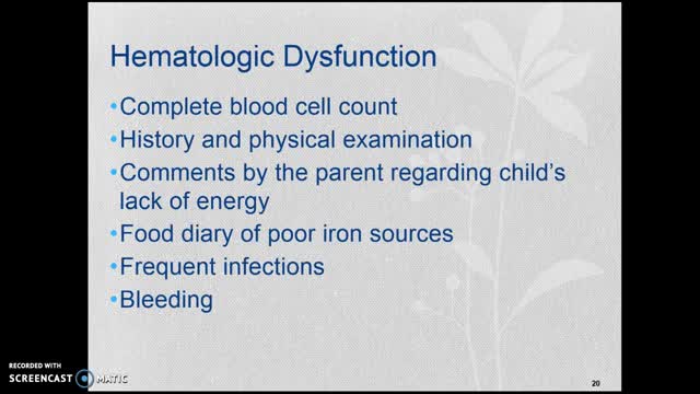
- Hematology
- Rheumatology
- Toxicology
- Nuclear Medicine
- Infectious Diseases
- Vascular Disease
- Reproductive Health
- Burns and Wound Healing
- Other
Latest videos

Folic acid, which is also called folate, is a B vitamin. The best food sources of folic acid are fortified cereals. Folic acid plays an important role in the production of red blood cells and helps your baby's neural tube develop into her brain and spinal cord.
Are you getting enough vitamin B12? Many people don’t, and that deficiency can cause some serious problems. Vitamin B12 does a lot of things for your body. It helps make your DNA and your red blood cells, for examples. Since your body doesn't make vitamin B12, you'll need to get it from animal-based foods or from supplements, and it needs to be consumed on a regular basis. Exactly how much you need and where you should get it from depends on things like your age, the diet you follow, your medical conditions, and in some cases what medications you take.

One of the most common parasites to infect human beings is the yeast-like Blastocystis hominis, a single-celled parasitic organism that causes abdominal cramping, bloating, gas, and sometimes anal itching. Other common parasites are: Tapeworms, which can grow as long as 60 feet while living in the human intestines.

Do you need to do a parasite cleanse? Probably... I hear from so many people suffering from symptoms of parasites - severe bloating, cramps, constipation, diarrhoea. A big problem in getting to the bottom of this (pun intended) is that the mainstream medical system really doesn’t have a way to detect, or even find most forms of parasites. They give you drugs for the symptoms, but essentially the parasites aren’t removed during that process.

Iron is a mineral that plays a vital role in health and well-being. Without it, many bodily functions would malfunction. “The primary role of iron is to carry oxygen in the blood to every cell in the body,” says Beth Thayer, RDN, MS, director of the Center for Health Promotion and Disease Prevention at Henry Ford Health System in Detroit. Iron is an important component of hemoglobin, the protein in red blood cells that carries oxygen from the lungs and transports it throughout the body.
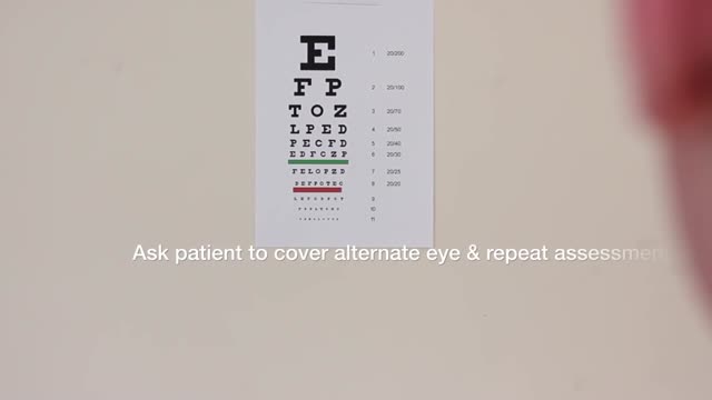
Examination of the Eyes and Vision
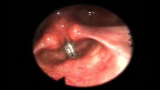
Throat Endoscopy: This video shows the vocal cords while singing
A lumbar puncture (also called a spinal tap) is a procedure to collect and look at the fluid (cerebrospinal fluid, or CSF) surrounding the brain and spinal cord. During a lumbar puncture, a needle is carefully inserted into the spinal canal low in the back (lumbar area). Samples of CSF are collected.
Malaria is a serious and sometimes fatal disease caused by a parasite that commonly infects a certain type of mosquito which feeds on humans. People who get malaria are typically very sick with high fevers, shaking chills, and flu-like illness.
Sickle cell anemia is an inherited form of anemia: This is a condition in which there aren't enough healthy red blood cells to carry adequate oxygen throughout your body. Normally, your red blood cells are flexible and round, moving easily through your blood vessels. In sickle cell anemia, the red blood cells become rigid and sticky and are shaped like sickles or crescent moons. These irregularly shaped cells can get stuck in small blood vessels, which can slow or block blood flow and oxygen to parts of the body. There's no cure for most people with sickle cell anemia. However, treatments can relieve pain and help prevent further problems associated with sickle cell anemia.
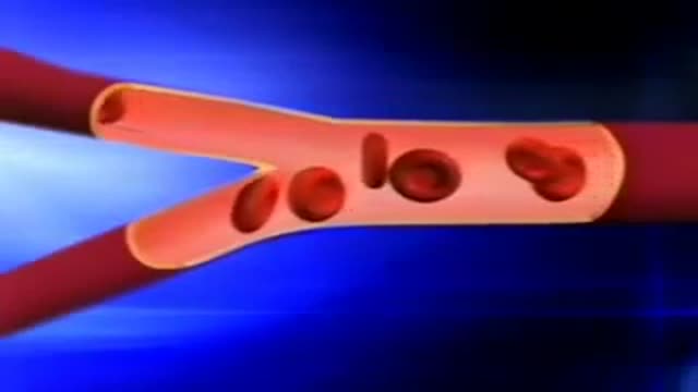
This video: Sickle cell anemia is an inherited form of anemia which is a condition in which there aren't enough healthy red blood cells to carry adequate oxygen throughout your body. Normally, your red blood cells are flexible and round, moving easily through your blood vessels. In sickle cell anemia, the red blood cells become rigid and sticky and are shaped like sickles or crescent moons. These irregularly shaped cells can get stuck in small blood vessels, which can slow or block blood flow and oxygen to parts of the body. There's no cure for most people with sickle cell anemia. However, treatments can relieve pain and help prevent further problems associated with sickle cell anemia.

Sickle cell anemia causes pain, fatigue and delayed growth, all because of a lack of enough healthy red blood cells. And yet genetic mutations that cause it — recessive genes for the oxygen-carrying hemoglobin protein — have survived natural selection because they also seem to provide a natural defense against malaria.
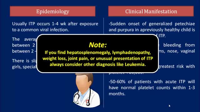
Idiopathic thrombocytopenic purpura (ITP) is a disorder that can lead to easy or excessive bruising and bleeding. The bleeding results from unusually low levels of platelets — the cells that help blood clot. Idiopathic thrombocytopenic purpura, which is also called immune thrombocytopenia, affects children and adults. Children often develop ITP after a viral infection and usually recover fully without treatment. In adults, the disorder is often long term. If you don't have signs of bleeding and your platelet count isn't too low, you may not need any treatment. In rare cases, the number of platelets may be so low that dangerous internal bleeding occurs. Treatment options are available.
Thrombotic thrombocytopenic purpura (TTP) is a rare blood disorder characterized by clotting in small blood vessels of the body (thromboses), resulting in a low platelet count. In its full-blown form, the disease consists of the pentad of microangiopathic hemolytic anemia, thrombocytopenic purpura, neurologic abnormalities, fever, and renal disease
TPE removes large-molecular-weight substances such as harmful antibodies from the plasma. It is usually carried out using an automated blood cell separator to ensure fluid balance and maintain a normal plasma volume. This may require the insertion of a femoral or jugular line to allow adequate blood flow. Typically, 30–40 mL/kg of plasma (1–1.5 plasma volumes) are removed at each procedure and replaced with isotonic 4.5 or 5.0% human albumin solution (some services substitute 25–50% of replacement volume with 0.9% saline). Exchange with fresh frozen plasma (FFP) is reserved for the replacement of ADAMTS13 in thrombotic thrombocytopenic purpura (see below) or to replace clotting factors. A one plasma volume exchange removes about 66% of an intravascular constituent and a two plasma volume exchange approximately 85%. TPE is normally combined with disease modifying treatment, such as immunosuppressive drugs, for the underlying condition.

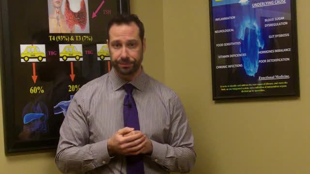
A thyroid biopsy is a procedure in which a small sample of tissue is removed from the thyroid gland and looked at under a microscope for cancer, infection, or other thyroid problems. The thyroid gland is found in front of the windpipe (trachea), just below the voice box (larynx). A sample of thyroid tissue can be taken by: Fine-needle biopsy. Your doctor puts a thin needle through the skin and into the thyroid gland. Many thyroid specialists like to use a needle biopsy method rather than surgery. Open biopsy. Your doctor makes a cut (incision) through the skin to see the thyroid gland. This method is done when other tests have not found the cause of your symptoms. Core needle biopsy. Your doctor inserts a needle with a special tip and removes a sample of tissue about the size of a grain of rice.
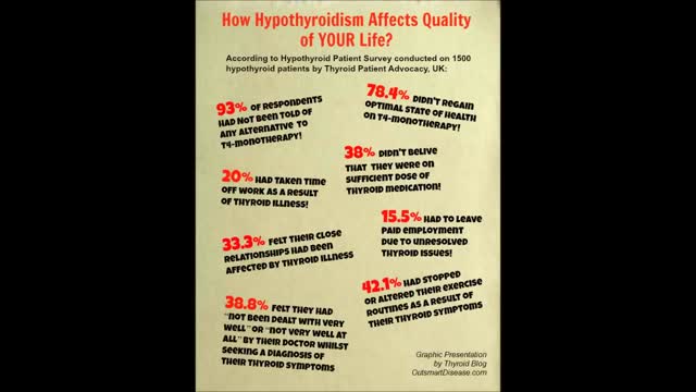
Through the hormones it produces, the thyroid gland influences almost all of the metabolic processes in your body. Thyroid disorders can range from a small, harmless goiter (enlarged gland) that needs no treatment to life-threatening cancer. The most common thyroid problems involve abnormal production of thyroid hormones. Too much thyroid hormone results in a condition known as hyperthyroidism. Insufficient hormone production leads to hypothyroidism. Although the effects can be unpleasant or uncomfortable, most thyroid problems can be managed well if properly diagnosed and treated.
A renal biopsy is a procedure used to extract kidney tissue for laboratory analysis. The word “renal” describes the kidneys. A renal biopsy is also called a kidney biopsy. The test helps your doctor identify the type of kidney disease you have, how severe it is, and the best treatment for it.
The kidneys are a pair of organs located in the back of the abdomen. Each kidney is about 4 or 5 inches long -- about the size of a fist. The kidneys' function are to filter the blood. All the blood in our bodies passes through the kidneys several times a day. The kidneys remove wastes, control the body's fluid balance, and regulate the balance of electrolytes. As the kidneys filter blood, they create urine, which collects in the kidneys' pelvis -- funnel-shaped structures that drain down tubes called ureters to the bladder. Each kidney contains around a million units called nephrons, each of which is a microscopic filter for blood. It's possible to lose as much as 90% of kidney function without experiencing any symptoms or problems.

Lumbar puncture is a common emergency department procedure used to obtain information about the cerebrospinal fluid (CSF) for diagnostic and, less commonly, therapeutic reasons. Please refer to the full article on Lumbar Puncture for more details on the lumbar puncture procedure. Lumbar puncture is typically performed via “blind” surface landmark guidance. The surface landmark technique is reported to be successful in a high percentage of attempted lumbar punctures; however, surface landmark identification of underlying structures has been shown to be accurate only 30% of the time. [1] Unsuccessful identification of proper landmarks often leads to increased difficulty in obtaining CSF, if the procedure is performed, and a higher rate of complications. Few alternatives are available in these cases. If available, fluoroscopic-guided lumbar puncture may be performed. If not, treatment is sometimes initiated empirically without obtaining CSF. Disadvantages of using fluoroscopy include limited availability or necessary transport of the patient outside of the emergency department, inability to directly visualize the spinal canal, and inherent radiation exposure
