Latest videos

This video: Blisters caused by friction or minor burns do not require a doctor's care. New skin will form underneath the affected area and the fluid is simply absorbed. Do not puncture a blister unless it is large, painful, or likely to be further irritated. The fluid-filled blister keeps the underlying skin clean, which prevents infection and promotes healing.

Most blisters caused by friction or minor burns do not require a doctor's care. New skin will form underneath the affected area and the fluid is simply absorbed. Do not puncture a blister unless it is large, painful, or likely to be further irritated. The fluid-filled blister keeps the underlying skin clean, which prevents infection and promotes healing.

Good and Bad Foods to Eat

Diverticula are small, bulging pouches that can form in the lining of your digestive system. They are found most often in the lower part of the large intestine (colon). Diverticula are common, especially after age 40, and seldom cause problems. Sometimes, however, one or more of the pouches become inflamed or infected. That condition is known as diverticulitis (die-vur-tik-yoo-LIE-tis). Diverticulitis can cause severe abdominal pain, fever, nausea and a marked change in your bowel habits. Mild diverticulitis can be treated with rest, changes in your diet and antibiotics. Severe or recurring diverticulitis may require surgery.

Initial symptoms may include: Pain or discomfort in the upper tummy (abdomen), especially after eating. Indigestion. (Note: most people who have indigestion do not have stomach cancer.) Feeling sick, and being off food. ... Weight loss and/or loss of appetite. You may pass blood out with your stools (faeces).
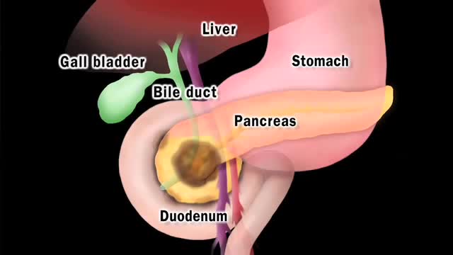
Pancreatic cancer begins in the tissues of your pancreas — an organ in your abdomen that lies horizontally behind the lower part of your stomach. Your pancreas secretes enzymes that aid digestion and hormones that help regulate the metabolism of sugars. Pancreatic cancer often has a poor prognosis, even when diagnosed early. Pancreatic cancer typically spreads rapidly and is seldom detected in its early stages, which is a major reason why it's a leading cause of cancer death. Signs and symptoms may not appear until pancreatic cancer is quite advanced and complete surgical removal isn't possible.

This video: Pancreatic cancer begins in the tissues of your pancreas — an organ in your abdomen that lies horizontally behind the lower part of your stomach. Your pancreas secretes enzymes that aid digestion and hormones that help regulate the metabolism of sugars. Pancreatic cancer often has a poor prognosis, even when diagnosed early. Pancreatic cancer typically spreads rapidly and is seldom detected in its early stages, which is a major reason why it's a leading cause of cancer death. Signs and symptoms may not appear until pancreatic cancer is quite advanced and complete surgical removal isn't possible.
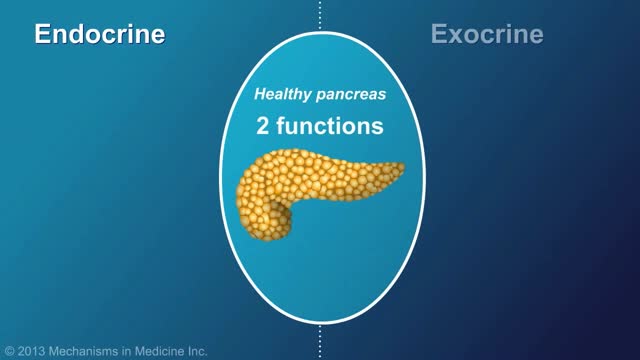
Chronic pancreatitis is a long-standing inflammation of the pancreas that alters the organ's normal structure and functions. It can present as episodes of acute inflammation in a previously injured pancreas, or as chronic damage with persistent pain or malabsorption.
Glomerular filtration is the first step in making urine. It is the process that your kidneys use to filter excess fluid and waste products out of the blood into the urine collecting tubules of the kidney, so they may be eliminated from your body.

If a patient comes to you with a painful, throbbing, swollen, red face (a ''fat face'), perhaps with fever, trismus and lymphadenitis, he is probably suffering from an acute dental or oral infection, most probably an alveolar abscess. He may have: (1) An alveolar abscess begins as an infection in the bone around a non-vital infected tooth. He has severe pain, which becomes less as pus is released into more superficial tissues and his face starts to swell. After 36 hours of cellulitis he usually has a fluctuant abscess which needs draining. If drainage is delayed, the pus in his abscess discharges spontaneously through a sinus (26-8) in his gum or face, which may become chronic. First, control infection with antibiotics, and then drain the abscess, either by incising it where it is pointing, or by removing the infected tooth, which acts as a cork to prevent the pus escaping, or by doing both these things. If you remove a tooth before you have controlled the infection with antibiotics, and while his face is still severely swollen, you may spread the infection; your task will also be more difficult. (2) A periodontal abscess at the side of a tooth, caused by spread from an infected gum. (3) A pericoronal abscess caused by infection of the gum over the crown of an unerupted and impacted tooth, usually a lower third molar (''an infected wisdom tooth'). Often, an abscess does not form, and the gum round the tooth is merely inflamed.

Red eyes usually are caused by allergy, eye fatigue, over-wearing contact lenses or common eye infections such as pink eye (conjunctivitis). However, redness of the eye sometimes can signal a more serious eye condition or disease, such as uveitis or glaucoma.

Exercises. Light exercises in which you move your affected limb may encourage lymph fluid drainage and help prepare you for everyday tasks, such as carrying groceries. ...
Doctors have many options to choose from, including interferon (Avonex, Betaseron, Extavia, and Rebif ), glatiramer acetate (Copaxone), mitoxantrone (Novantrone), teriflunomide (Aubagio), fingolimod (Gilenya), dimethyl fumarate (Tecfidera), and natalizumab (Tysabri).

http://destructeur-de-poids.info-pro.co Perdre Des Cuisses, Astuces Pour Maigrir, Objectif Ventre Plat, Exercice Pour Perdre Du Poids. Boire de l’eau est relié au gain de poids Ceci peut sembler vraiment étrange... Mais boire de l'eau serait la raison pourquoi la plupart d'entre nous avons des problèmes à perdre du poids ? Une découverte récente démontre que c'est vrai et que pour perdre du poids... il faut connaître exactement la quantité d'eau à boire... basé sur votre propre corps. En utilisant ce truc simple, les gens ont perdu 15, 20, 25 kilos même, en moins de 2 mois. Découvrez combien d'eau vous avez besoin et commencez à perdre du poids. http://destructeur-de-poids.info-pro.co http://astuce-ventre-plat.blogspot.com/
Angina is a term used for chest pain caused by reduced blood flow to the heart muscle. Angina (an-JIE-nuh or AN-juh-nuh) is a symptom of coronary artery disease. Angina is typically described as squeezing, pressure, heaviness, tightness or pain in your chest. Angina, also called angina pectoris, can be a recurring problem or a sudden, acute health concern. Angina is relatively common but can be hard to distinguish from other types of chest pain, such as the pain or discomfort of indigestion. If you have unexplained chest pain, seek medical attention right away.
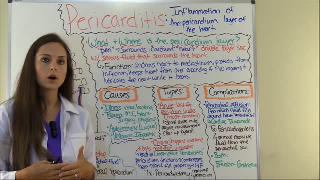
Pericarditis is an inflammation of the lining surrounding the heart (the pericardial sac). Pericardial effusion is a collection of fluid in the pericardial sac. This fluid may be produced by inflammation. The cause of pericarditis in most individuals is unknown but is likely due to viral infection.

Best Sex Position to Get Pregnant Fast

Vaginismus is unique because it may result from a combination of physical or non-physical causes—or seem to have none at all.

SEX WITH DIAPHRAGM TO CONTROL UNWANTED PREGNANCY
female condom
