Latest videos
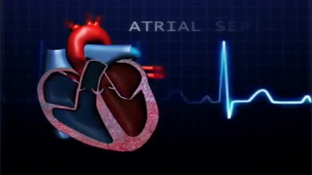
atrial septal defect (ASD) is a hole in the wall between the two upper chambers of your heart (atria). The condition is present from birth (congenital). Small atrial septal defects may close on their own during infancy or early childhood. Large and long-standing atrial septal defects can damage your heart and lungs. Small defects may never cause a problem and may be found incidentally. An adult who has had an undetected atrial septal defect for decades may have a shortened life span from heart failure or high blood pressure that affects the arteries in the lungs (pulmonary hypertension). Surgery may be necessary to repair atrial septal defects to prevent complications
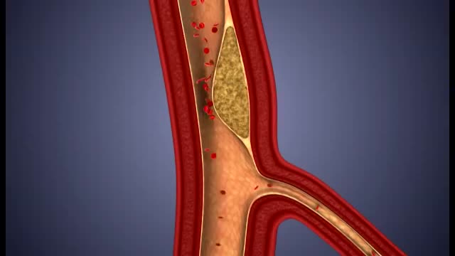
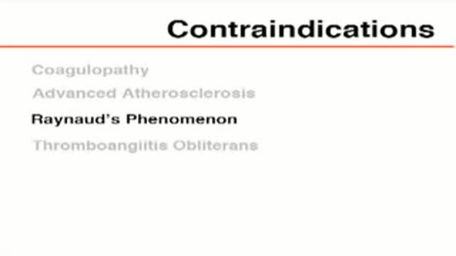
What damage does atherosclerosis cause? Plaque may partially or totally block the blood's flow through an artery in the heart, brain, pelvis, legs, arms or kidneys. Some of the diseases that may develop as a result of atherosclerosis include coronary heart disease, angina (chest pain), carotid artery disease, peripheral artery disease (PAD) and chronic kidney disease.
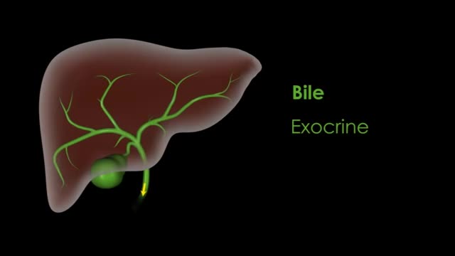
liver also detoxifies chemicals and metabolizes drugs. As it does so, the liver secretes bile that ends up back in the intestines. The liver also makes proteins important for blood clotting and other functions.

It is a specialized bodily fluid that supplies essential substances and nutrients, such as sugar, oxygen, and hormones to our cells, and carries waste away from those cells, this waste is eventually flushed out of the body in urine, feces, sweat, and lungs (carbon dioxide). Blood also contains clotting agents.

Blood type (or blood group) is determined, in part, by the ABO blood group antigens present on red blood cells. A blood type (also called a blood group) is a classification of blood based on the presence or absence of inherited antigenic substances on the surface of red blood cells (RBCs).

Blood cells travel through the circulatory system suspended in a yellowish fluid called plasma. Plasma is 90% water and contains nutrients, proteins, hormones, and waste products. Whole blood is a mixture of blood cells and plasma.

Life Before Birth
Fake Sonogram
A central venous catheter, also called a central line, is a long, thin, flexible tube used to give medicines, fluids, nutrients, or blood products over a long period of time, usually several weeks or more. A catheter is often inserted in the arm or chest through the skin into a large vein.
There are two main purposes of an arterial line. Firstly when patients are very sick an arterial line is inserted to provide constant monitoring and recording of the patient's blood pressure. Secondly some patients require frequent blood tests and the arterial line provides easy access to a patient's blood.
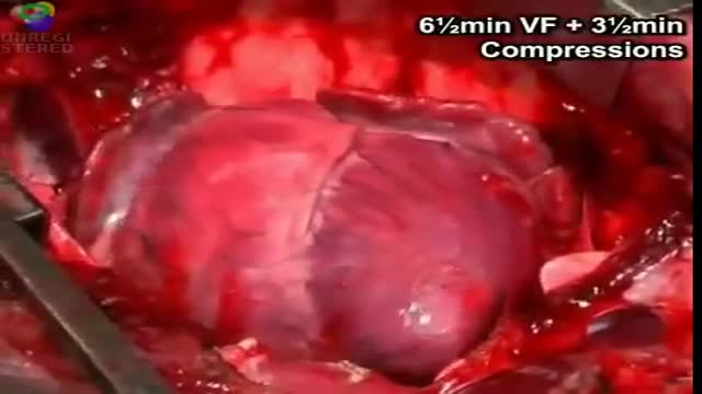
cardiac massage intermittent compression of the heart by pressure applied either over the sternum (closed cardiac massage) or directly to the heart through an opening in the chest wall (open cardiac massage). simple massage in the nursing interventions classification, a nursing intervention defined as stimulation of the skin and underlying tissues with varying degrees of hand pressure to decrease pain, produce relaxation, and/or improve circulation.
Stapling is used to treat prolapsed hemorrhoids. A surgical staple fixes the prolapsed hemorrhoid back into place inside your rectum and cuts off the blood supply so that the tissue will shrink and be reabsorbed. Stapling recovery takes less time and is less painful than recovery from a hemorrhoidectomy.
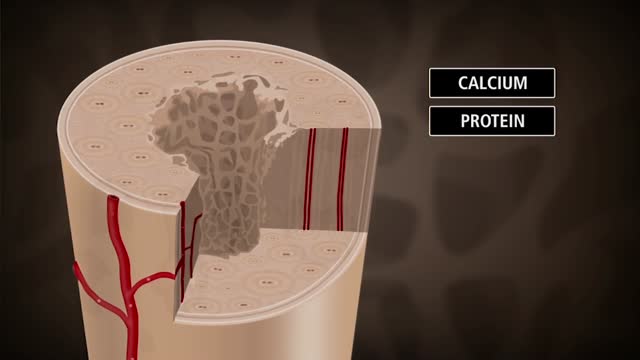
Bone is not a static part of the body — it's constantly being resorbed (broken down) and formed throughout your life. Your entire skeleton is replaced about every decade, according to the NIH. During your childhood and teenage years, bone formation occurs more quickly than bone resorption, resulting in growth. You reach your maximum bone density and strength around age 30, after which bone resorption slowly overtakes bone formation. Osteoporosis develops when there's an abnormal imbalance between bone resorption and formation — that is, resorption occurs too quickly, or formation too slowly.
Osteoporosis is a disease in which bones become brittle and fragile due to low bone mass and bone tissue loss. It's the most common type of bone disease, according to the National Institutes of Health (NIH), and increases your risk of fractures, particularly of the hips, spine, and wrists. Prevalence In the United States, nearly 54 million people ages 50 and older were living with osteoporosis or osteopenia (low bone mass ) in 2010, according to a 2014 article in the Journal of Bone and Mineral Research. More specifically, 10.2 million adults had osteoporosis, and 43.4 million adults had osteopenia, which puts a person at high risk for osteoporosis.
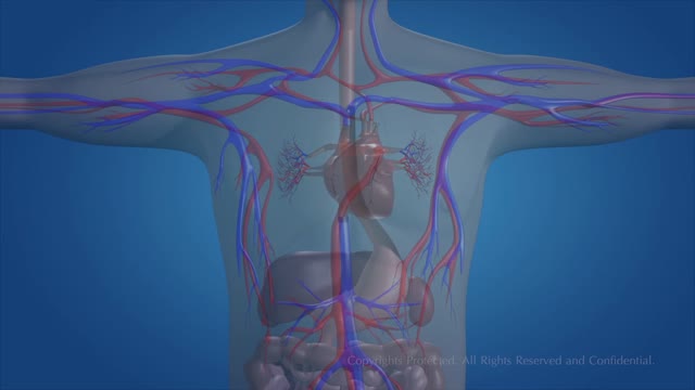
The essential components of the human cardiovascular system are the heart, blood and blood vessels. It includes the pulmonary circulation, a "loop" through the lungs where blood is oxygenated; and the systemic circulation, a "loop" through the rest of the body to provide oxygenated blood.
Premature ventricular contractions (PVCs) are extra, abnormal heartbeats that begin in one of your heart's two lower pumping chambers (ventricles). These extra beats disrupt your regular heart rhythm, sometimes causing you to feel a flip-flop or skipped beat in your chest.
An intra-aortic balloon pump (IABP) is a mechanical device that helps the heart pump blood. This device is inserted into the aorta, the body's largest artery. It is a long, thin tube called a catheter with a balloon on the end of it. If you are hospitalized, your doctor may insert an IABP.

Dextrocardia situs inversus refers to the heart being a mirror image situated on the right side. For all visceral organs to be mirrored, the correct term is dextrocardia situs inversus totalis. Dextrocardia is believed to occur in approximately 1 in 12,000 people.
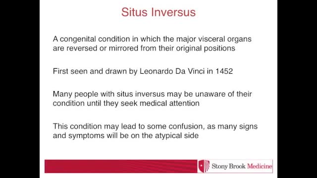
Situs inversus is a condition in which the arrangement of the internal organs is a mirror image of normal anatomy. It can occur alone (isolated, with no other abnormalities or conditions) or it can occur as part of a syndrome with various other defects. Congenital heart defects are present in about 5-10% of affected people. The underlying cause and genetics of situs inversus are complex. Familial cases have been reported.[1][2]
The term hypoplastic left heart syndrome (HLHS), initially proposed by Noonan and Nadas, [1] describes a spectrum of cardiac abnormalities characterized by marked hypoplasia of the left ventricle and ascending aorta. This is the same disorder characterized as hypoplasia of the aortic tract complex by Lev. [2] The aortic and mitral valves are atretic, hypoplastic, or stenotic. A patent foramen ovale or an atrial septal defect is usually present. The ventricular septum is usually intact. A large patent ductus arteriosus supplies blood to the systemic circulation. Systemic arterial desaturation may be present because of complete mixing of pulmonary and systemic venous blood in the right atrium. Coarctation of the aorta is also commonly present. See the images below.
