- Physical Examination
- Surgical Examination
- Ophthalmology
- Clinical Skills
- Orthopedics
- Surgery Videos
- Laparoscopy
- Pediatrics
- Funny Videos
- Cardiothoracic Surgery
- Nursing Videos
- Plastic Surgery
- Otorhinolaryngology
- Histology and Histopathology
- Neurosurgery
- Dermatology
- Pediatric Surgery
- Urology
- Dentistry
- Oncology and Cancers
- Anatomy Videos
- Health and Fitness
- Radiology
- Anaesthesia
- Physical Therapy
- Pharmacology
- Interventional Radiology
- Cardiology
- Endocrinology
- Gynecology
- Emergency Medicine
- Psychiatry and Psychology
- Childbirth Videos
- General Medical Videos
- Nephrology
- Physiology
- Diet and Food Health
- Diabetes Mellitus
- Neurology
- Women Health
- Osteoporosis
- Gastroenterology
- Pulmonology
- Hematology
- Rheumatology
- Toxicology
- Nuclear Medicine
- Infectious Diseases
- Vascular Disease
- Reproductive Health
- Burns and Wound Healing
- Other
Latest videos
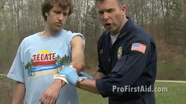
how to control arterial bleeding Learn more at http://www.ProTrainings.com
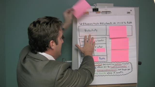
Why chiropractic us crucial to your health
How we went from super species to the sickest. What gravity has to do with it.

Mitral Valve Repair video

Insulina, Sintomi Iperglicemia, Dolci Per Diabetici Ricette, Giornata Mondiale Del Diabete
http://diabete-cura.info-pro.co
Abbassa il livello di zucchero nel sangue e liberati dall’insulina in tre settimane o meno ... GARANTITO!
Ogni giorno, negli Stati Uniti, vengono diagnosticati più di 2000 nuovi casi di diabete.
Con l'attenzione focalizzata sui livelli di zucchero nel sangue e insulina, tuttavia, la causa di tutta la devastazione è stata trascurata.
Questo ebook rivoluzionario rivela la causa principale del diabete e come invertire il processo.
Il diabete è una malattia che se non si prende un'azione efficace contro di essa, peggiora sempre di più.
Purtroppo, i farmaci curano solo i sintomi del diabete e di solito non fanno nulla per affrontare le cause sottostanti.
PER COMINCIARE A GUARIRE DAL DIABETE
come curare il diabete in modo naturale e garantito senza farmaci e insulina! CLICCA QUI: http://diabete-cura.info-pro.co
Clicca sul link sottostante per fare il check out
http://diabete-cura.info-pro.co
Iscriviti al nostro canale
http://www.youtube.com/user/viveresano01
https://www.youtube.com/watch?v=1sul1papqgk
Insulina, Sintomi Iperglicemia, Dolci Per Diabetici Ricette, Giornata Mondiale Del Diabete,
diabete mellito,
sintomi diabete,
diabete sintomi,
diabete tipo 2,
dieta per diabetici,
diabete insipido,
diabete gestazionale,
diabete tipo 1,
dolci per diabetici,
sintomi del diabete,
iperglicemia,
diabete mellito tipo 2,
dieta diabete

Stay active and push your body to its limit – tips on how you can mend strained muscles and prevent injury.
How to keep those creepy crawly lice from pestering your family’s scalps– tips on prevention and removal.
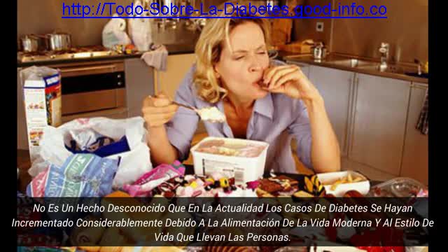
Causas De La Diabetes, Signos De La Diabetes, Complicaciones Agudas De La Diabetes, Diabetico
http://todo-sobre-la-diabetes.good-info.co
Remedios Naturales Para Controlar La Diabetes
No es un hecho desconocido que en la actualidad los casos de diabetes se hayan incrementado considerablemente debido a la alimentación de la vida moderna y al estilo de vida que llevan las personas.
La diabetes tipo 2 es una enfermedad que se relaciona profundamente con la alimentación y se caracteriza por un elevado nivel de azúcar en sangre.
Este tipo de diabetes se puede controlar perfectamente llevando un estilo de vida saludable y una alimentación apropiada.
Existen muchos remedios naturales por los que puedes optar para luchar contra la diabetes:
Se trata de un remedio natural muy sencillo de realizar y que te resultará de gran utilidad para combatir la diabetes.
como eliminar la diabetes en pocos dias de manera natural y para siempre haciendo click aqui:
http://todo-sobre-la-diabetes.good-info.co
Haga Clic En El Enlace De Abajo Para Comprobar Que Funciona
http://todo-sobre-la-diabetes.good-info.co
Suscríbete A Nuestro Canal
https://www.youtube.com/user/VivirConSalud1
https://www.youtube.com/watch?v=i89z59Oi7Bg
Causas De La Diabetes, Signos De La Diabetes, Complicaciones Agudas De La Diabetes, Diabetico,
tipos de diabetes que existen,
Causa De La Diabetes,
que provoca la diabetes,
que ocasiona la diabetes,
historia natural de la diabetes mellitus,
fisiopatologia del pie diabetico,
cuales son las causas de la diabetes,
como se detecta la diabetes,
como detectar la diabetes,
clasificacion de la diabetes,
federacion internacional de diabetes
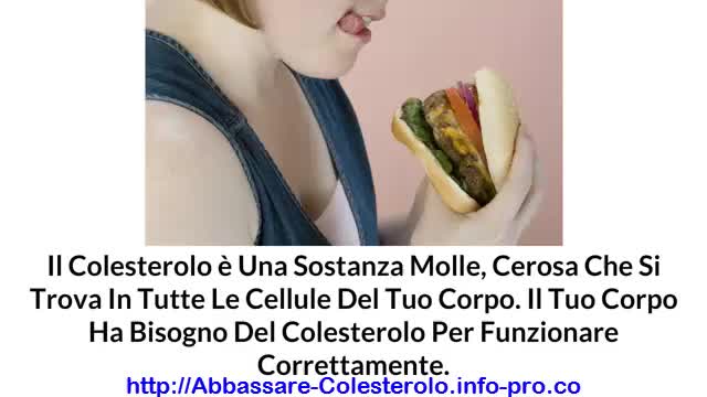
Come Alzare Il Colesterolo Buono, Colesterolo Hdl, Abbassare Il Colesterolo
http://abbassare-colesterolo.info-pro.co
COME EFFICACEMENTE ABBASSARE IL COLESTEROLO
senza prendere farmaci!
Il colesterolo è una sostanza molle, cerosa che si trova in tutte le cellule del tuo corpo. Il tuo corpo ha bisogno del colesterolo per funzionare correttamente. Il tuo corpo utilizza il colesterolo per tenere insieme le cellule. Inoltre il tuo corpo usa il colesterolo per creare gli ormoni, la vitamina D, e sostanze che aiutano a digerire gli alimenti.
Tuttavia, se troppo colesterolo entra nel sangue può causare problemi. Questo è noto come il colesterolo alto.
Se hai il colesterolo alto, e non fai nulla per abbassarlo, sarai ad un maggior rischio di gravi problemi di salute, come ad esempio un attacco di cuore o ictus. Pertanto, l'abbassamento del colesterolo è una questione importante per la salute generale di tutti.
Per saperne di più su come si può seguire un piano scientificamente provato per sconfiggere il colesterolo, visita il sito:
http://abbassare-colesterolo.info-pro.co
Clicca sul link sottostante per fare il check out
http://abbassare-colesterolo.info-pro.co
Iscriviti al nostro canale
http://www.youtube.com/user/viveresano01
https://www.youtube.com/watch?v=BWojp9nfdsU
Come Alzare Il Colesterolo Buono, Colesterolo Hdl, Abbassare Il Colesterolo,
colesterolo,
alimenti colesterolo,
colesterolo ldl basso,
colesterolo alimenti da evitare,
colesterolo e trigliceridi alti,
alimenti contro il colesterolo,
dieta x colesterolo,
cause colesterolo alto,
cosa mangiare per abbassare il colesterolo,
alimenti ricchi di colesterolo,
cibi contro il colesterolo
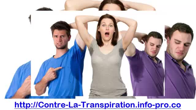
Transpire, Stop Transpiration, Transpiration Des Aisselles, Probleme Transpiration, Transpirer Moins
http://contre-la-transpiration.info-pro.co
La transpiration des aisselles
La transpiration est à la fois positive et négative pour le corps humain.
Ce processus est à l’origine de l’élimination de toxines présentes dans l’organisme. Mais parfois certains sont gênés par les traces de sueur et l’odeur attachée à leurs vêtements.
Heureusement, il existe de nombreux moyen d’empêcher la transpiration des aisselles.
Des méthodes et des produits naturels peuvent se révéler tout aussi efficaces que des interventions chirurgicales.
La première mesure à prendre pour limiter la transpiration des aisselles est d’être attentif à votre hygiène.
Dès que vous sentez que vos aisselles sont humides, lavez-les avec un savon hypoallergénique et essuyez-les doucement avec un linge doux.
Certains antitranspirants contenant 20% de ce composant sont disponibles en vente libre.
Ainsi votre corps fonctionnera en harmonie car il sera mieux équilibré sur le plan hormonal.
Enfin! Découvrez Une Méthode Simple Pour Lutter contre la Transpiration
Cliquez ici: http://contre-la-transpiration.info-pro.co
Cliquez Sur Le Lien Ci-dessous Pour Vérifier
http://contre-la-transpiration.info-pro.co
Abonnez-vous à Notre Chaîne
https://www.youtube.com/user/AideADomicile11
https://www.youtube.com/watch?v=KpymjEtVsiM
Transpire, Stop Transpiration, Transpiration Des Aisselles, Probleme Transpiration, Transpirer Moins,
comment lutter contre la transpiration,
vetement anti transpiration,
chaussettes anti transpiration,
déodorant anti transpirant efficace,
je transpire,
anti transpirant homme,
je transpire des pieds,
ne plus transpirer,
transpiration excessive dos,
transpiration jaune,
probleme de transpiration aisselle
Hair transplant is a life-altering decision. If you are worried about hair loss, or consider baldness a hindrance, then you are ready to take the next step. Now the question is what to do next? Obviously, the worst choice would be to do "nothing" at all! Secondly, you could try to preserve your existing hair with medicines, remedies and hair-care products - it might just work for you. Thirdly, you could go for a hair-piece or a wig. But if you're reading this, then the chances are that you're looking for a permanent solution for your hair problem, which can best be provided through a hair transplant -an increasingly popular method of defeating baldness and patchy hair.
http://www.cocoona.ae/hair_transplantation.asp

If you are unhappy with the shape and contours of your breast, then you are not alone. Millions of women around the world are unhappy with their breasts either because they are too small or too big, or too distorted.
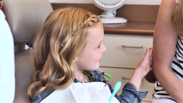
Your kids are going to love brushing! Follow these tips and find out how brushing your teeth can be fun and effective for the whole family.

Comment Avoir Un Ventre Plat, Rajeuni, Rajeunir De 10 Ans En 3 Mois, Bruleur De Graisse Musculation
http://rajeunir-de-10-ans.info-pro.co
5 Raisons pourquoi le Cardio Long-Lent n’est PAS bon.
En passant à travers les e-mails de clients au cours des derniers jours, j'ai remarqué que beaucoup de gens font encore du cardio longue durée, à faible intensité, beurk!
Voici l'affaire: si vous cherchez à obtenir un bénéfice maximal du temps que vous mettez dans vos séances d'entraînement, le cardio de longue durée à faible intensité n'est pas la voie à suivre, et pour de nombreuses raisons.
En fait, je n’ai le temps pour faire que quelques heures d'exercice par semaine, et vous savez quoi? C'est tout ce dont vous avez besoin. En fait, la recherche a montré que plus de 90 minutes par semaine peuvent être nuisibles! (Plus d'infos ici)
Ces 5 étapes Révèlent Les Choses Que Vous Devez ABSOLUMENT ÉVITER Si Vous Voulez Ralentir Le Processus De Vieillissement, Récupérer Votre Santé Et Atteindre Un Corps Idéal.
Cliquez Ici: http://rajeunir-de-10-ans.info-pro.co
Cliquez Sur Le Lien Ci-dessous Pour Vérifier
http://rajeunir-de-10-ans.info-pro.co
Abonnez-vous à Notre Chaîne
https://www.youtube.com/user/AideADomicile11
https://www.youtube.com/watch?v=ja2ygliMwjA
Comment Avoir Un Ventre Plat, Rajeuni, Rajeunir De 10 Ans En 3 Mois, Bruleur De Graisse Musculation,
comment brûler les graisses abdominales,
programme perdre du poids,
ventre plat homme,
brules graisse,
qui brûle,
des conseils pour perdre du poids,
bruleur de graisse naturelle,
bruler les graisse du ventre,
programme brule graisse,
programme alimentaire pour perdre du poids,
regime alimentaire perdre du poids,
brulle graisse,
juvamine brule graisse
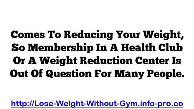
How To Lose Weight In A Week, How To Lose Fat Without Exercise, Diets That Work Fast
http://lose-weight-without-gym.info-pro.co
3 Ways For Fast Weight Loss
There are many people who would like to reduce their weight very fast sitting at home; cost is a constraint when it comes to reducing your weight, so membership in a health club or a weight reduction center is out of question for many people.
The ways of weight loss at home fast is not a rocket science exertion rather it’s like an open book. The most important rules are taking a balanced diet which is rich in fiber, cutting of carbohydrates from the meals and planning out exercises and work outs properly.
The following section discusses these 3 simple yet effective ways of weight loss.
How to train your brain to lose pounds
Click Here: http://lose-weight-without-gym.info-pro.co
Click The Link Below To Check It Out
http://lose-weight-without-gym.info-pro.co
Subscribe To Our Channel
https://www.youtube.com/user/NaturalFatBurners1
https://www.youtube.com/watch?v=EcicJ5J-C7U
How To Lose Weight In A Week, How To Lose Fat Without Exercise, Diets That Work Fast,
gym workout to lose weight,
how to lose weight in gym,
losing weight at the gym,
best workout for losing weight,
best gym workout to lose weight,
how to lose weight permanently,
how can you lose weight in a week,
great exercises to lose weight,
what is the best workout to lose weight,
what are the best exercises to lose weight
How smoking is causing a hazy filter over your life. Kick cigarettes to the curb for good.
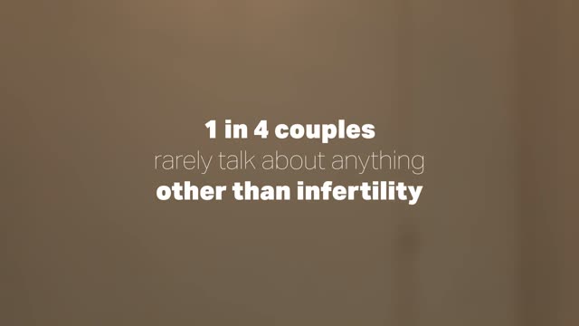
The impacts of infertility from the bedroom to the bank account – options to support the journey to parenthood.
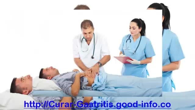
Remedios Caseros Para La Gastritis, Remedio Para La Gastritis, Jugos Para La Gastritis
http://curar-gastritis.good-info.co
La cura natural comienza desde el interior revirtiendo las causas que la originan.
“La gastritis” se da por problemas digestivos y la deficiente función del sistema inmunológico, resultado de una incorrecta alimentación y de malos hábitos.
Se debe curar la raíz, “La Raíz Son Los Hábitos En El Estilo De Vida”, de lo contrario nada efectivo sucederá por más antiácidos, antibióticos o simples remedios caseros que tomemos. E incluso ni una cirugía la soluciona. Al contrario todo esto complica el problema.
La cura natural y definitiva se logra aplicando un conjunto de técnicas como las siguientes:
. Incorporando una correcta nutrición con alimento físico y otros elementos importantes de la naturaleza.
. Incorporando hábitos saludables.
. Infusión de hierbas.
. El consumo de minerales naturales.
. Evitando el consumo alimentos tóxicos y dañinos.
. Eliminando el consumo de medicamentos.
La verdad De Cómo Eliminar La Gastritis De Raíz
Con El Método 100% Natural Y El Mas Efectivo Que Existe
Haciendo Click Aqui: http://curar-gastritis.good-info.co
Haga Clic En El Enlace De Abajo Para Comprobar Que Funciona
http://curar-gastritis.good-info.co
Suscríbete A Nuestro Canal
https://www.youtube.com/user/VivirConSalud1
https://www.youtube.com/watch?v=g61B6C6m2Cg
Remedios Caseros Para La Gastritis, Remedio Para La Gastritis, Jugos Para La Gastritis,
consecuencias de la gastritis,
dieta para personas con gastritis,
que es la gastritis cronica,
sintomas de gastritis aguda,
tratamiento natural para la gastritis,
sintomas de la gastritis cronica,
remedio casero para gastritis,
alimentos para gastritis,
sintomas del helicobacter pylori,
gastritis cie 10,
helicobacter pylori síntomas,
remedio para gastritis
Pastillas Para La Ereccion, Problemas De Ereccion Por Nervios, Como Tener Mas Ereccion
http://erecciones-fuertes-duraderas.good-info.co
La Solución Natural Para Alcanzar La Mejor Performance Sexual.
No es extraño escuchar que los hombres gastan cada vez más dinero para someterse a tratamientos para recuperar y mantener la erección de su órgano viril, tratamientos que además de caros suelen provocar numerosos daños a la salud.
Sin embargo, usted puede revertir su disfunción sexual modificando algunos de sus hábitos de vida. De lo que se trata es de hacer cambios simples y posibles, todos aquellos cambios que serán el mejor remedio natural para combatir la disfunción eréctil.
Los cambios simples y posibles darán resultados en breve tiempo. El deseo sexual y la erección dejarán de verse frustrados a poco de comenzar a llevar a cabo las recomendaciones.
El Único Sistema Efectivo Y Natural Para Combatir La Disfunción Eréctil
Haciendo Click Aqui: http://erecciones-fuertes-duraderas.good-info.co
Haga Clic En El Enlace De Abajo Para Comprobar Que Funciona
http://erecciones-fuertes-duraderas.good-info.co
Suscríbete A Nuestro Canal
https://www.youtube.com/user/VivirConSalud1
https://www.youtube.com/watch?v=djpiGp8j70A
Pastillas Para La Ereccion, Problemas De Ereccion Por Nervios, Como Tener Mas Ereccion,
falta de ereccion,
que es impotencia,
problemas de ereccion soluciones,
problemas de ereccion,
remedios para la impotencia masculina,
tengo problemas de ereccion,
remedios para impotencia,
remedios para impotencia masculina,
ereccion masculina,
problemas de ereccion causas,
fumar causa impotencia
Tudo Sobre Diabetes, Diabetes Tem Cura, O Que é Diabetes Tipo 2, Plantas Que Curam Diabetes
http://tudo-sobre-diabetes.good-info.co
Cura Naturalmente a Diabetes Tipo 2
A diabetes tipo II se tornou uma das doenças mais comuns nos tempos modernos. A boa notícia é que em pouco menos de um mês, seguindo um plano de alimentação e vida saudável, é possível equilibrar seu nível de açúcar no sangue e prevenir as terríveis consequências que esta doença tem.
A seguir, você encontrará este plano para nivelar o açúcar no sangue e dizer adeus para a diabetes.
Restrinja o consumo de todo o tipo de bebidas.
Realize atividade física de baixo impacto todo o dia, por um mínimo de meia hora.
Elimine por completo de suas refeições, todos os alimentos que contenham farinha branca.
Inclua em sua alimentação habitual, ácidos gordos essenciais (especialmente ácidos ômega 3), inclua também o consumo de frutas secas.
único Sistema Eficiente, Fácil E Natural Para Eliminar Para Sempre O Diabetes. Um Sistema Cientificamente Comprovado
Clique No Link Abaixo Para Verificá-la
http://tudo-sobre-diabetes.good-info.co
Assine O Nosso Canal
https://www.youtube.com/user/dicasdesaude11
https://www.youtube.com/watch?v=61MN7xSR9yA
Tudo Sobre Diabetes, Diabetes Tem Cura, O Que é Diabetes Tipo 2, Plantas Que Curam Diabetes,
diabetes gestacional,
diabetes mellitus tipo 2,
diabetes dieta,
sintomas de diabete,
diabetes tipo 1 e 2,
medicamentos para diabetes,
diabete sintomas,
causas da diabetes,
como evitar diabetes,
sintomas da diabetes tipo 2,
tratamento da diabetes,
o que diabetes,
os sintomas da diabete
