- Physical Examination
- Surgical Examination
- Ophthalmology
- Clinical Skills
- Orthopedics
- Surgery Videos
- Laparoscopy
- Pediatrics
- Funny Videos
- Cardiothoracic Surgery
- Nursing Videos
- Plastic Surgery
- Otorhinolaryngology
- Histology and Histopathology
- Neurosurgery
- Dermatology
- Pediatric Surgery
- Urology
- Dentistry
- Oncology and Cancers
- Anatomy Videos
- Health and Fitness
- Radiology
- Anaesthesia
- Physical Therapy
- Pharmacology
- Interventional Radiology
- Cardiology
- Endocrinology
- Gynecology
- Emergency Medicine
- Psychiatry and Psychology
- Childbirth Videos
- General Medical Videos
- Nephrology
- Physiology
- Diet and Food Health
- Diabetes Mellitus
- Neurology
- Women Health
- Osteoporosis
- Gastroenterology
- Pulmonology
- Hematology
- Rheumatology
- Toxicology
- Nuclear Medicine
- Infectious Diseases
- Vascular Disease
- Reproductive Health
- Burns and Wound Healing
- Other
Latest videos

http://mylapbandsuccess.plus101.com
---Lap Band Success Stories And Pictures. "My Journey With The Lap Band Has Been Rough... In Fact, I Almost Took Matters Into My Own Hands, And Ended That Journey A few Years Ago...
But the fact that I'm still here today -- slim and happy, is proof that it is possible to overcome, and end your emotional reliance on food, and get the body and life you've always wanted!
Let Me Tell You My Story...
It was the worst week I had since my surgery -- Thanksgiving week. I was at a point where all I was thinking about all day, was food. I had to actually fight to resist my strong cravings...
I just wanted to take one bite of that turkey... just eat one piece of pumpkin pie! I just wanted to be able to taste some of the same things everyone else was eating!
Not Only Was I Still Obese, But I Couldn't Even Enjoy My Own Life!
My cravings were driving me insane, but I did my best to resist the temptation. When I got onto the scales that week, I was dumb-founded - My weight loss had stalled! All the agony and deprivation I'd suffered... was for nothing!
I was so angry at myself for even putting me in such a low, pitiful state in the first place! I was to blame for the way I looked! I was frustrated and... I felt helpless!
My Weight Loss Had Stalled...
Ok, so maybe I needed another refill but nothing could improve my emotional state-of-mind...
I became so disillusioned that I could not remember my reasonsfor wanting to lose weight, and how critical it was for me to resist my favorite foods. All I thought, was that the lap band was not working for me... and I gave in!
I Was Once Too Embarrassed To Share My Story With Anyone, But I'm Telling It To You Today, So You'll Know That You're Not Alone!...
I had lost all hope of losing weight, that I began to out-eat my band, and find ways to cheat it...
One of my favorite foods before the surgery was french fries dipped in a mix of mayonnaise and ketchup. Since I couldn't have it after the lap band, I improvised... and blended it!
I Would Actually Put Fries, Mayo And Ketchup Into A Blender... And Then Drink It!
If you've ever fallen off the wagon then I don't have to tell you about theguilt that sets in after-wards...
I'd drink it, then I'd feel guilty and start to cry...
It would make me sick... but I did it anyway!
I hated myself for doing it... but I did it anyway!
When the scales began to creep back up again, I knew why... but I didn't know what to do about it!
I felt like my life was in a tail-spin. The worst part was that I was just too embarrassed & humiliated to talk to anyone about it!
Have you ever felt that way?
To find out what happened next, scroll down!
In an amazing twist of fate, find out how I found it easier to lose the last 132 lbs than it ever was to lose the first 60 lbs!"
By April Cannon
Lap, Band, Success, Stories, And, Pictures, Laparoscopic, gastric, Banding, costs, stomach, surgery, diet, successfull, Raisa Khan, April Cannon
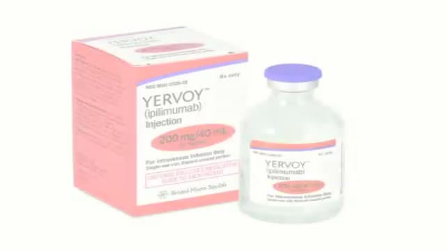
How revolutionizing advancements helps patients with metastatic melanoma kick start the body’s immune system to increase survival.
Inguinal hernia repair without mesh, Desarda Repair, no recurrence, no pain, no mesh hernia surgery, hernia operation, no mesh, without mesh, hernia operation, hernia surgery, new method
In the video, Dr Hilary Jones talks about the important role the gut has in the immune system and the valuable role second generation prebiotics such as Bimuno IMMUNAID can play.
The winter season is upon us and with it comes the dreaded flu season. Some people are more susceptible than others and so, it’s important to be aware that not only does the gut play a central role in your immune system but there are positive measures we can take to support it. In this video, Dr Hilary Jones talks about the important role the gut has in the immune system and the valuable role second generation prebiotics can play.
Bimuno IMMUNAID RRP £9.99 for 30 pastilles. Available from Boots and www.bimuno.com. Find us on Facebook and follow us on twitter @BimunoUK
How ESC therapy rejuvenates our body?

How ESC therapy treats diseases?
ESC therapy technology in China since 2006

ESC therapy helps in treatment of aging problems, degenrative diseases, heart & kidney failures, spinal cord injury, parkinson's , alzheimer, diabetes etc.
Treatment for Piles,Fistula,hemorrhoids, Hydrocele Without Operation or surgery in pakistan Dr Jamil Ahmad Hashmi ( haripur hazar pakistan )... +923009885511 --- drjamil79@gmail.com
Treatment for Piles,Fistula,Hydrocele Without Operation piles treatment with 60 days Quickly! pain free treatment full life Piles Medicine dr jamil ahmad hashmi ( haripur hazar pakistan ) drjamil79@yahoo.com +923009885511 piles treatment with 60 days Quickly! pain free treatment full life Piles Medicine dr jamil ahmad hashmi...
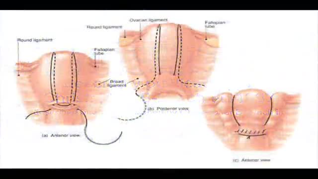
B-Lynch suture for uterine atony technique described
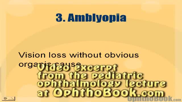
How amblyopia develops in children. Basically, if one eye doesn't see well from an early age, the wiring never forms correctly back to the occipital cortex.
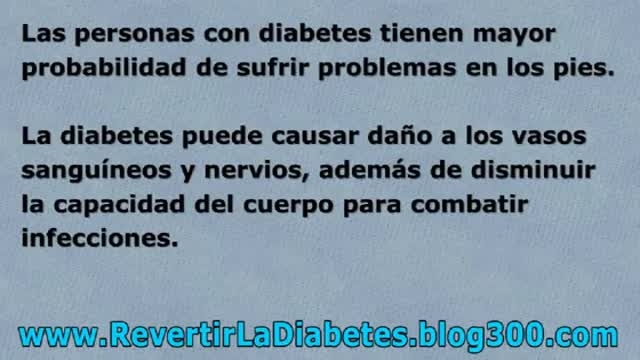
http://revertirlaDiabetes.blog300.com - Como Se Cura La Diabetes - Como Tratar la Diabetes - Como Prevenir La Diabetes
Aprenda Cómo Vencer La Diabetes y Recuperar Su Salud
Te sugiero conocer un metodo que ayudó finalmente a muchas personas
que padecian de problemas de Diabetes
Ingresa AQUI
http://revertirlaDiabetes.blog300.com
Como Se Cura La Diabetes - Como Tratar la Diabetes - Como Prevenir La Diabetes
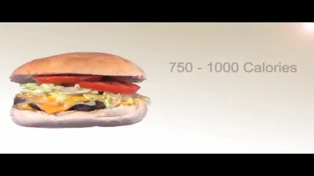
With healthy ingredients in the comfort of your home in no time flat
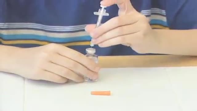
With a portable pump controlled by a wireless handheld device that automatically delivers insulin.
alternative ingredients for healthy meals and diabetes management.
The menopause experience is different for everyone so explore options to manage symptoms from proper diet and exercise to hormone therapy.

From fabric choices to layering, clothing tips to help you deal with menopausal hot flashes
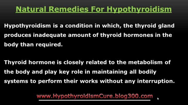
http://www.HypothyroidismCure.blog300.com - Hypothyroidism Diet Plan - Hypothyroidism Treatment - Hypothyroidism Revolution
10 Hypothyroidism Diet Tips to Help Heal Your Thyroid
1. Avoid Anti-Thyroid Foods
2. Increase Your Saturated Fats
3. Eat Your Fruit
4. Increase Your Salt Intake
5. Get Plenty of Bone Broth
6. Eat Some Shellfish
7. Cut the Processed Foods
8. Cook Your Veggies
9. Don’t Overdo the Water
10. Drink Your Coffee
Hypothyroidism Diet Plan - Hypothyroidism Treatment - Hypothyroidism Revolution

http://www.HypothyroidismCure.blog300.com - Hypothyroidism Treatment Natural - Hypothyroidism Recipes Treatment
Let’s Get Something Straight…
* You’re here because you’re serious about overcoming your hypothyroidism…
* You’re here because you’re serious about and taking back your life…
* You know there’s no magic pill to cure your hypothyroidism and never will be…
Hypothyroidism Treatment Natural - Hypothyroidism Recipes Treatment
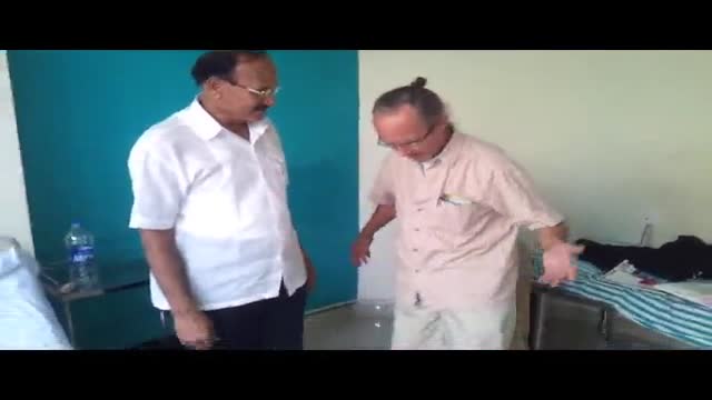
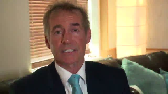
Considering having an Austin plastic surgery procedure like Smartlipo? Then you’ll want to watch this quick video where staff members of renowned Austin plastic surgeon Dr. William Davis give you an overview of what you can expect.
