- Physical Examination
- Surgical Examination
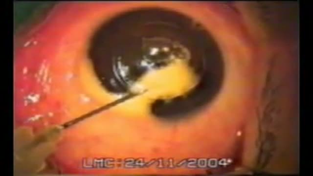
- Ophthalmology
- Clinical Skills
- Orthopedics
- Surgery Videos
- Laparoscopy
- Pediatrics
- Funny Videos
- Cardiothoracic Surgery
- Nursing Videos
- Plastic Surgery
- Otorhinolaryngology
- Histology and Histopathology
- Neurosurgery
- Dermatology
- Pediatric Surgery
- Urology
- Dentistry
- Oncology and Cancers
- Anatomy Videos
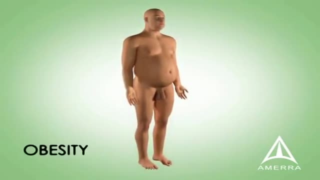
- Health and Fitness
- Radiology
- Anaesthesia
- Physical Therapy
- Pharmacology
- Interventional Radiology
- Cardiology
- Endocrinology
- Gynecology
- Emergency Medicine
- Psychiatry and Psychology
- Childbirth Videos
- General Medical Videos
- Nephrology
- Physiology
- Diet and Food Health
- Diabetes Mellitus
- Neurology
- Women Health
- Osteoporosis
- Gastroenterology
- Pulmonology
- Hematology
- Rheumatology
- Toxicology
- Nuclear Medicine
- Infectious Diseases
- Vascular Disease
- Reproductive Health
- Burns and Wound Healing
- Other
Latest videos
The Role of Insulin in the Human Body
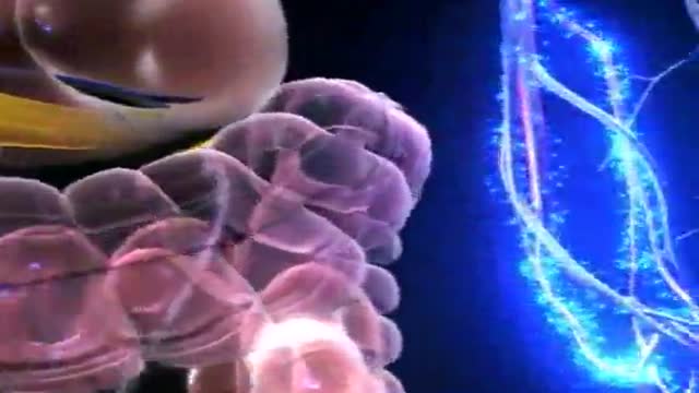
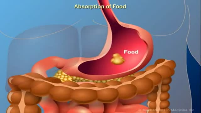
Diabetes Animation 3D
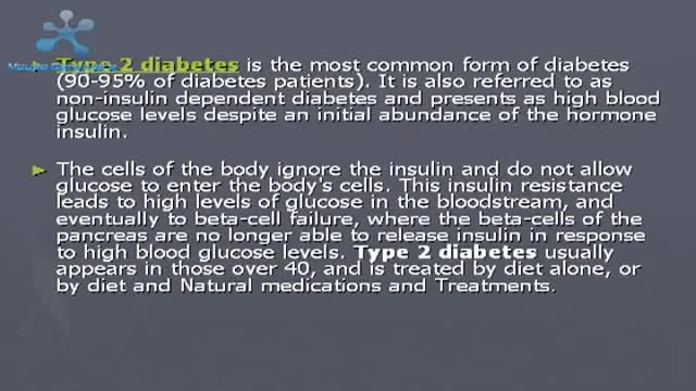
Type 2 Diabetes Causes Symptoms and Treatment
How to Reverse Type 2 Diabetes
How to Know If You Have Diabetes
Type 2 Diabetes Animation 3D
Insulin Processes Mechanism Animation 3D
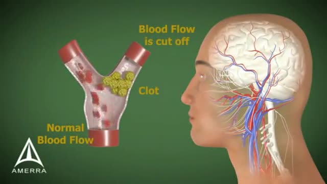
Stroke Animation 3D
Erectile Dysfunction Information 3D Animation
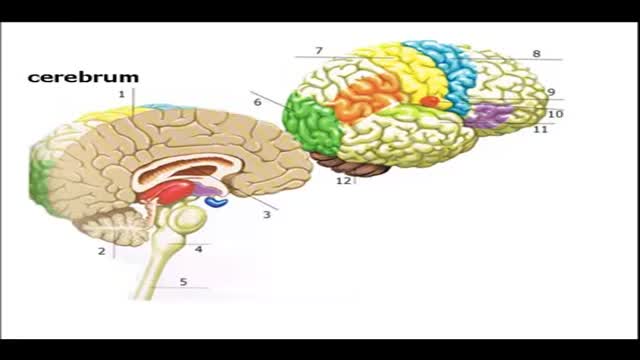
Brain Anatomy and Functions Animation

Wernicke's aphasia is a neurological disorder typically caused by stroke. It affects the Wernicke's region in the brain's left hemisphere which is reasoned to be responsible for processing of meaning, especially as it relates to verbal communication, hence the problems with speech witnessed in these patients
Big Bubble Technique

Robot Flies Like a Bird
Giving Birth
Sutureless laparoscopic radical Prostatectomy

Woman Giving Birth
Birth

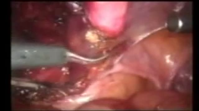
Bilateral Laparoscopic Pyeloplasty in Children

Delivery Video
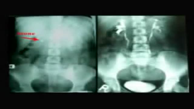
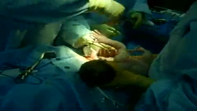
Totally US Guided PCNL in Flank Position
