Pinakabagong mga video
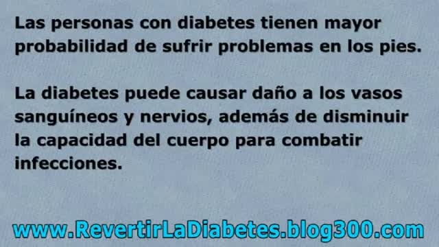
http://revertirlaDiabetes.blog300.com - Como Se Cura La Diabetes - Como Tratar la Diabetes - Como Prevenir La Diabetes
Aprenda Cómo Vencer La Diabetes y Recuperar Su Salud
Te sugiero conocer un metodo que ayudó finalmente a muchas personas
que padecian de problemas de Diabetes
Ingresa AQUI
http://revertirlaDiabetes.blog300.com
Como Se Cura La Diabetes - Como Tratar la Diabetes - Como Prevenir La Diabetes
With healthy ingredients in the comfort of your home in no time flat
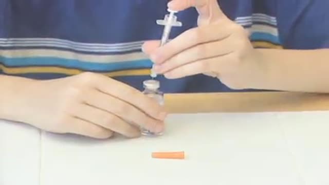
With a portable pump controlled by a wireless handheld device that automatically delivers insulin.
alternative ingredients for healthy meals and diabetes management.

The menopause experience is different for everyone so explore options to manage symptoms from proper diet and exercise to hormone therapy.

From fabric choices to layering, clothing tips to help you deal with menopausal hot flashes
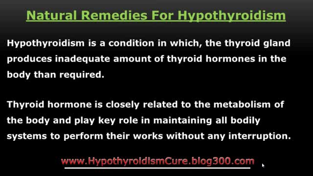
http://www.HypothyroidismCure.blog300.com - Hypothyroidism Diet Plan - Hypothyroidism Treatment - Hypothyroidism Revolution
10 Hypothyroidism Diet Tips to Help Heal Your Thyroid
1. Avoid Anti-Thyroid Foods
2. Increase Your Saturated Fats
3. Eat Your Fruit
4. Increase Your Salt Intake
5. Get Plenty of Bone Broth
6. Eat Some Shellfish
7. Cut the Processed Foods
8. Cook Your Veggies
9. Don’t Overdo the Water
10. Drink Your Coffee
Hypothyroidism Diet Plan - Hypothyroidism Treatment - Hypothyroidism Revolution

http://www.HypothyroidismCure.blog300.com - Hypothyroidism Treatment Natural - Hypothyroidism Recipes Treatment
Let’s Get Something Straight…
* You’re here because you’re serious about overcoming your hypothyroidism…
* You’re here because you’re serious about and taking back your life…
* You know there’s no magic pill to cure your hypothyroidism and never will be…
Hypothyroidism Treatment Natural - Hypothyroidism Recipes Treatment
Considering having an Austin plastic surgery procedure like Smartlipo? Then you’ll want to watch this quick video where staff members of renowned Austin plastic surgeon Dr. William Davis give you an overview of what you can expect.
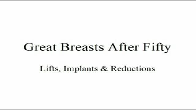
Plastic Surgery New York Dr. Carlin Vickery of 5th Avenue Aesthetics Surgery in Manhattan
(http://www.5thavesurgery.com) speaks at a Fab Over 50 event on having great breasts after the age
of 50. In this presentation Dr. Carlin shares patient results by providing before and after pictures from
different types of breast surgeries including breast lifts, implants and reductions.

Going to the dentist is not a very fun experience for most. In fact, let's face it, most of us dread it.
http://www.dentistmaps.com/
Brain Concussion Recognize and Report
Brain Concussion in Sports

Brain Concussion Animation
Brain Concussion Accidents Examples

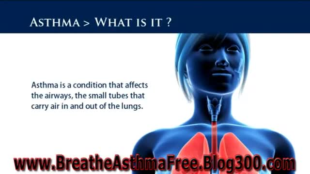
http://breatheasthmafree.blog300.com - Bronchitis Asthma Symptoms - Asthma Treatments For Adults
Asthma Sufferer?
Ex-Sufferer Reveals Own Natural
Remedy for Eliminating Asthma!...
http://breatheasthmafree.blog300.com
Bronchitis Asthma Symptoms - Asthma Treatments For Adults
http://breatheasthmafree.blog300.com - Asthma Treatments bronchitis - Bronchitis Asthma Home Remedies
Kill Asthma Today!
Natural - Treatment for Asthma
Cure & Revitalise Your Breathing
http://breatheasthmafree.blog300.com
Asthma Treatments bronchitis - Bronchitis Asthma Home Remedies
Dr Allen’s device provides a new kidney stones treatment that tackles the cause of kidney stone formation which is hidden at the capillary level, read at http://www.finetreatment.co.uk. The unique natural Thermobalancing Therapy does not use harmful medication or shock waves and, of course, surgery. Learn by watching this video about kidney stones cause and how to dissolve kidney stone or kidney stones at home by using Dr Allen’s natural therapeutic device.
The 30 minute DVD:
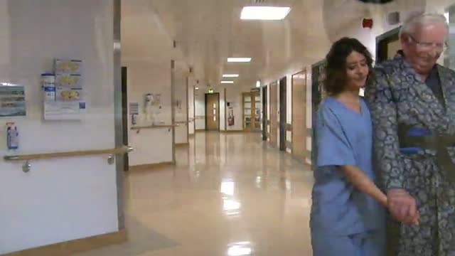
introduces moving and handling of people
describes safer people handling practices
features specialist guidance from a chartered physiotherapist
outlines the process for people handling risk assessments
sets out the principles of safer handling
demonstrates the key safer handling techniques:
rolling a person
inserting and removing sliding sheets
repositioning people using sliding sheets
assisting people to stand and walk with handling belts
the use of roll boards in lateral transfers
using hoists
highlights the important role you play in safer people handling
