- Physical Examination
- Surgical Examination
- Ophthalmology
- Clinical Skills
- Orthopedics
- Surgery Videos
- Laparoscopy
- Pediatrics
- Funny Videos
- Cardiothoracic Surgery
- Nursing Videos
- Plastic Surgery
- Otorhinolaryngology
- Histology and Histopathology
- Neurosurgery
- Dermatology
- Pediatric Surgery
- Urology
- Dentistry
- Oncology and Cancers
- Anatomy Videos
- Health and Fitness
- Radiology
- Anaesthesia
- Physical Therapy
- Pharmacology
- Interventional Radiology
- Cardiology
- Endocrinology
- Gynecology
- Emergency Medicine
- Psychiatry and Psychology
- Childbirth Videos
- General Medical Videos
- Nephrology
- Physiology
- Diet and Food Health
- Diabetes Mellitus
- Neurology
- Women Health
- Osteoporosis
- Gastroenterology
- Pulmonology
- Hematology
- Rheumatology
- Toxicology
- Nuclear Medicine
- Infectious Diseases
- Vascular Disease
- Reproductive Health
- Burns and Wound Healing
- Other
Latest videos

Angiogenesis and Cancer
Alicia Berger
7,879 Views • 2 years ago
Angiogenesis and Cancer
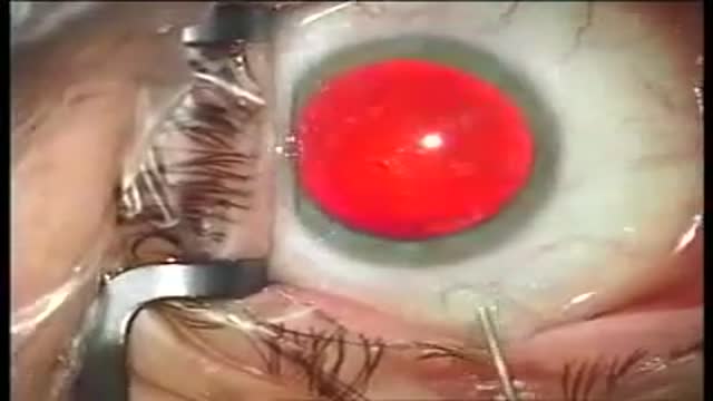
RLE and Alcon Restor IOL Implantation
Alicia Berger
8,119 Views • 2 years ago
RLE and Alcon Restor IOL Implantation
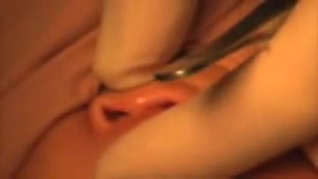
Cleft Lip Surgery Millard Unilateral
Alicia Berger
11,854 Views • 2 years ago
Cleft Lip Surgery Millard Unilateral

Immunization Mechanism Animation
Alicia Berger
1,484 Views • 2 years ago
Immunization Mechanism Animation

Mumps Signs Symptoms Complications
Alicia Berger
1,380 Views • 2 years ago
Mumps Signs Symptoms Complications
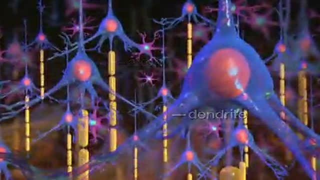
Alzheimer Disease Effects
Alicia Berger
1,720 Views • 2 years ago
Alzheimer Disease Effects

Lung Vasculature
Alicia Berger
11,624 Views • 2 years ago
Lung Vasculature

Urinary and Fecal Incontinence Animation
Alicia Berger
1,690 Views • 2 years ago
Urinary and Fecal Incontinence Animation

Emboli Formation in Artery
Alicia Berger
26,916 Views • 2 years ago
Emboli Formation in Artery

MRI of Fetal Brain Development
Alicia Berger
8,029 Views • 2 years ago
MRI of Fetal Brain Development
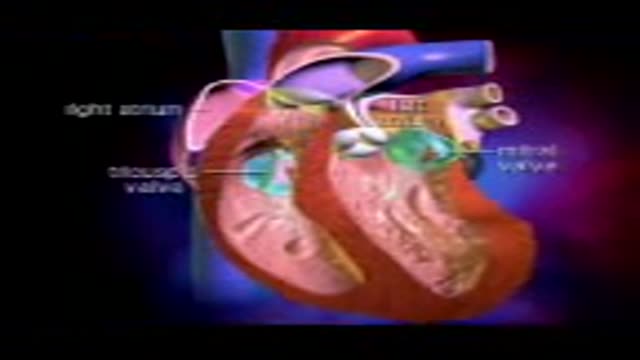
Heart Valves
Alicia Berger
2,138 Views • 2 years ago
Heart Valves

Gastrointestinal GI Drug Delivery
Alicia Berger
24,928 Views • 2 years ago
Gastrointestinal GI Drug Delivery
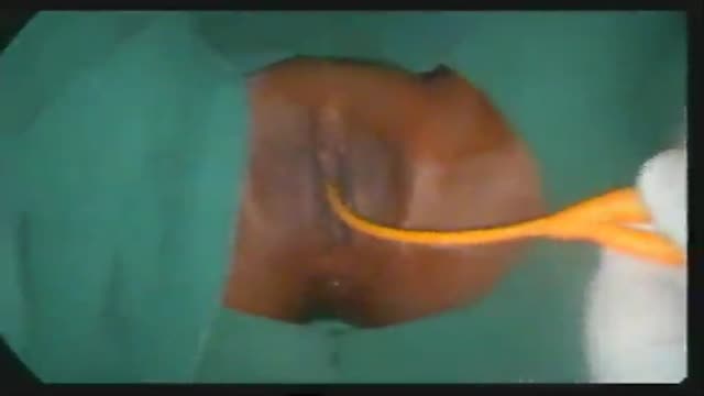
Imperforate Hymen
Alicia Berger
16,069 Views • 2 years ago
Imperforate Hymen

Lower Face Lift Awake Plastic Surgery
Alicia Berger
11,157 Views • 2 years ago
Lower Face Lift Awake Plastic Surgery
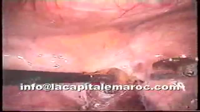
Pelvic lymphadenectomy
Alicia Berger
6,423 Views • 2 years ago
Pelvic lymphadenectomy
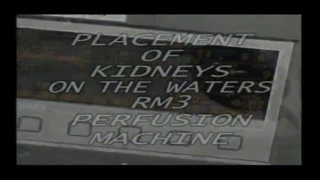
Dissection and Cannulation of Cadaveric Kidney
Alicia Berger
13,046 Views • 2 years ago
Dissection and Cannulation of Cadaveric Kidney
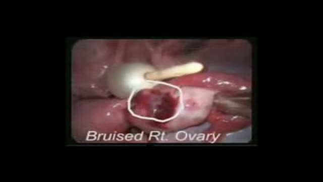
Traumatic Urinary Bladder Groin Injury
Alicia Berger
9,044 Views • 2 years ago
Traumatic Urinary Bladder Groin Injury
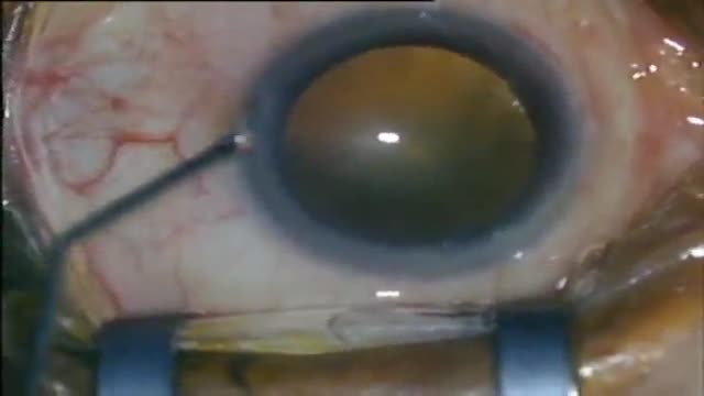
New Phacoemulsification Horizontal Chopping
Alicia Berger
6,903 Views • 2 years ago
New Phacoemulsification Horizontal Chopping
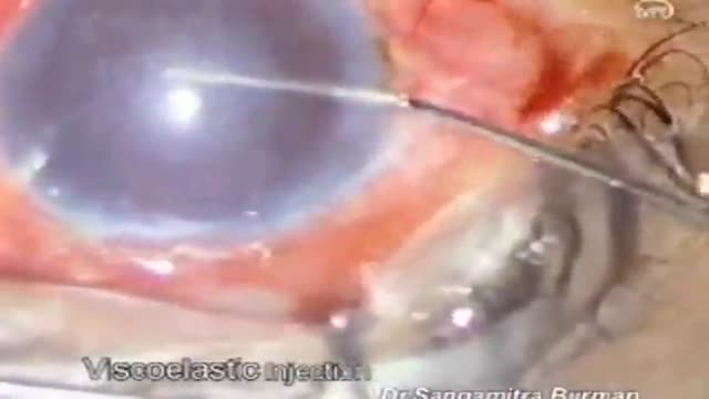
Eye Surgery penetrating Keratoplasty
Alicia Berger
10,710 Views • 2 years ago
Eye Surgery penetrating Keratoplasty
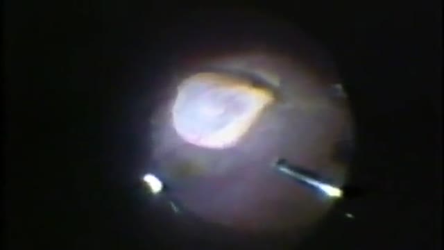
Eye Cyst Removal By Vitrectomy
Alicia Berger
6,785 Views • 2 years ago
Eye Cyst Removal By Vitrectomy
Showing 266 out of 267
