- Physical Examination
- Surgical Examination
- Ophthalmology
- Clinical Skills
- Orthopedics
- Surgery Videos
- Laparoscopy
- Pediatrics
- Funny Videos
- Cardiothoracic Surgery
- Nursing Videos
- Plastic Surgery
- Otorhinolaryngology
- Histology and Histopathology
- Neurosurgery
- Dermatology
- Pediatric Surgery
- Urology
- Dentistry
- Oncology and Cancers
- Anatomy Videos
- Health and Fitness
- Radiology
- Anaesthesia
- Physical Therapy
- Pharmacology
- Interventional Radiology
- Cardiology
- Endocrinology
- Gynecology
- Emergency Medicine
- Psychiatry and Psychology
- Childbirth Videos
- General Medical Videos
- Nephrology
- Physiology
- Diet and Food Health
- Diabetes Mellitus
- Neurology
- Women Health
- Osteoporosis
- Gastroenterology
- Pulmonology
- Hematology
- Rheumatology
- Toxicology
- Nuclear Medicine
- Infectious Diseases
- Vascular Disease
- Reproductive Health
- Burns and Wound Healing
- Other
Latest videos
Eye Surgery Trabeculectomy
Alicia Berger
7,914 Views • 2 years ago
Eye Surgery Trabeculectomy
Eye Surgery C Lasik
Alicia Berger
6,696 Views • 2 years ago
Eye Surgery C Lasik
Abnormal Eye Lid Positions Ptosis
Alicia Berger
6,555 Views • 2 years ago
Abnormal Eye Lid Positions Ptosis

Gastric Lavage Video
Alicia Berger
15,638 Views • 2 years ago
Gastric Lavage Video
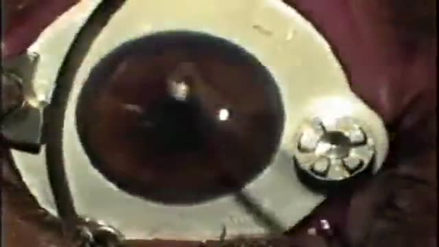
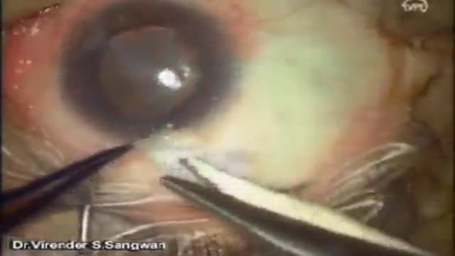
Eye Phacoemulsification
Alicia Berger
6,268 Views • 2 years ago
Eye Phacoemulsification
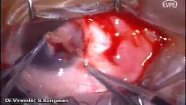
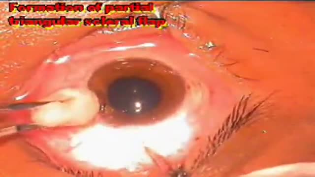
Pterygium Excision with Auto Conjunctival Graft
Alicia Berger
8,555 Views • 2 years ago
Pterygium Excision with Auto Conjunctival Graft
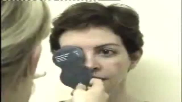
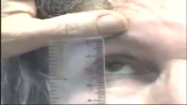
Ocular Movement Examination
Alicia Berger
7,055 Views • 2 years ago
Ocular Movement Examination
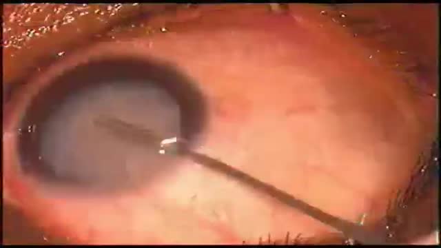
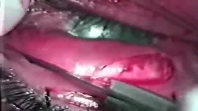
Keratectomy with Amniotic Membrane Graft Eye
Alicia Berger
7,137 Views • 2 years ago
Keratectomy with Amniotic Membrane Graft Eye
Eye Lid Tarsal Fracture Surgery
Alicia Berger
7,338 Views • 2 years ago
Eye Lid Tarsal Fracture Surgery
Eye Lid Partial Tarsectomy Surgery
Alicia Berger
6,099 Views • 2 years ago
Eye Lid Partial Tarsectomy Surgery
Eye Lid Jones Procedure
Alicia Berger
5,998 Views • 2 years ago
Eye Lid Jones Procedure
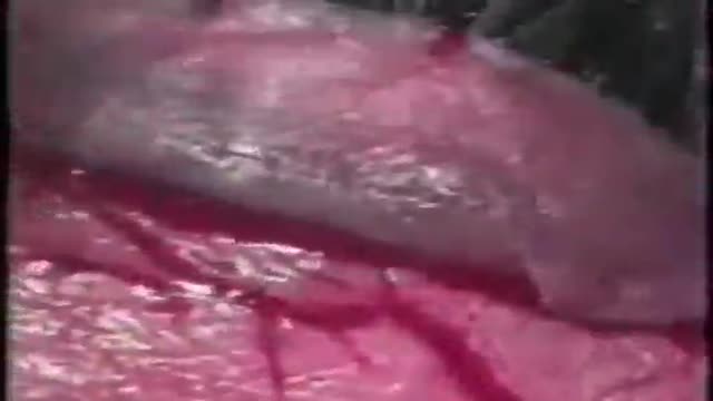
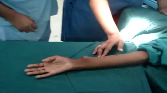
Arteriolotomy Open Heart Surgery
Alicia Berger
10,865 Views • 2 years ago
Arteriolotomy Open Heart Surgery
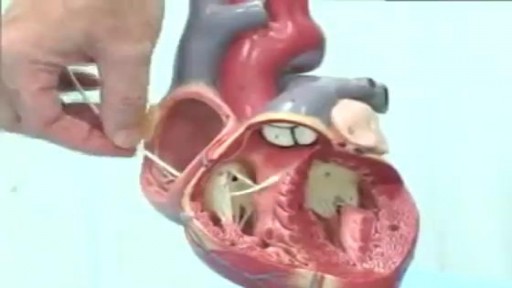
Pulmonary Artery Swan Ganz Catheter
Alicia Berger
9,478 Views • 2 years ago
Pulmonary Artery Swan Ganz Catheter
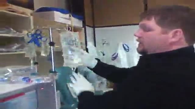
Hemodialysis Machine Setup
Alicia Berger
8,480 Views • 2 years ago
Hemodialysis Machine Setup
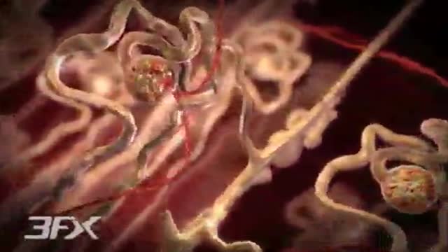
Diabetic Nephropathy Animation 3D
Alicia Berger
12,175 Views • 2 years ago
Diabetic Nephropathy Animation 3D
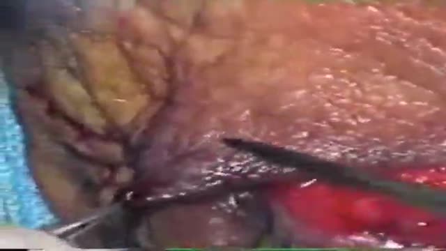
Ligation of Aneurysm in ArterioVenous Malformation
Alicia Berger
7,224 Views • 2 years ago
Ligation of Aneurysm in ArterioVenous Malformation
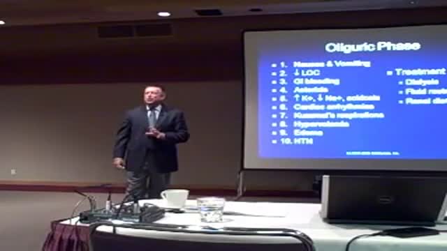
Acute Renal Failure for Nursing
Alicia Berger
11,294 Views • 2 years ago
Acute Renal Failure for Nursing
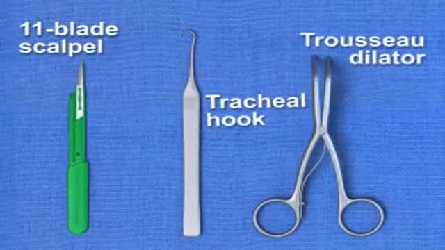
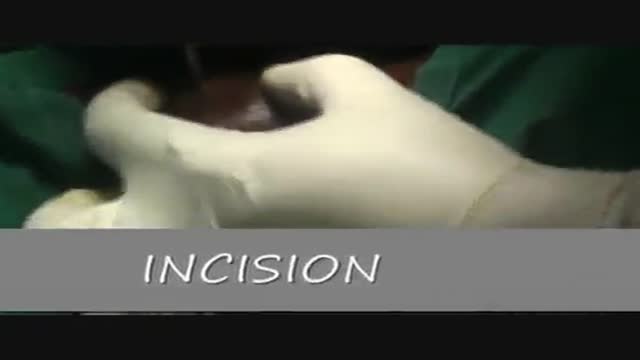
Traditional Surgical Cricothyrotomy
Alicia Berger
9,880 Views • 2 years ago
Traditional Surgical Cricothyrotomy
Cricothyrotomy Quick Airway Access
Alicia Berger
8,676 Views • 2 years ago
Cricothyrotomy Quick Airway Access
Mechanism of Type 2 Diabetes Animation
Alicia Berger
10,407 Views • 2 years ago
Mechanism of Type 2 Diabetes Animation
Showing 267 out of 268
