- Physical Examination
- Surgical Examination
- Ophthalmology
- Clinical Skills
- Orthopedics
- Surgery Videos
- Laparoscopy
- Pediatrics
- Funny Videos
- Cardiothoracic Surgery
- Nursing Videos
- Plastic Surgery
- Otorhinolaryngology
- Histology and Histopathology
- Neurosurgery
- Dermatology
- Pediatric Surgery
- Urology
- Dentistry
- Oncology and Cancers
- Anatomy Videos
- Health and Fitness
- Radiology
- Anaesthesia
- Physical Therapy
- Pharmacology
- Interventional Radiology
- Cardiology
- Endocrinology
- Gynecology
- Emergency Medicine
- Psychiatry and Psychology
- Childbirth Videos
- General Medical Videos
- Nephrology
- Physiology
- Diet and Food Health
- Diabetes Mellitus
- Neurology
- Women Health
- Osteoporosis
- Gastroenterology
- Pulmonology
- Hematology
- Rheumatology
- Toxicology
- Nuclear Medicine
- Infectious Diseases
- Vascular Disease
- Reproductive Health
- Burns and Wound Healing
- Other
Latest videos
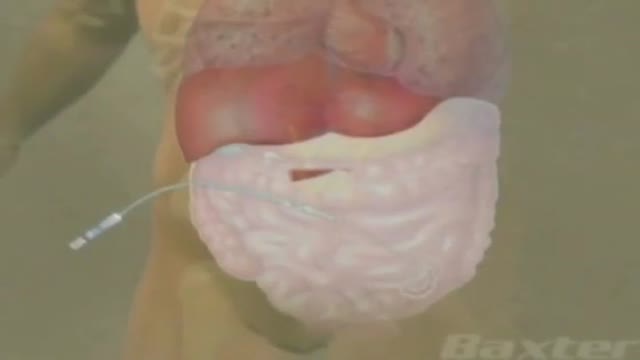
Peritoneal Dialysis for Kidney Disease

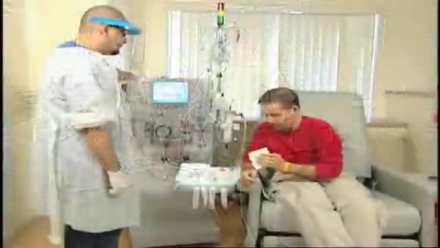
Hemodialysis Introduction for Kidney
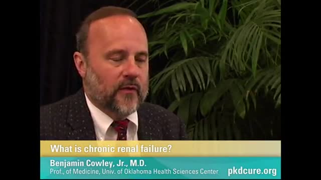
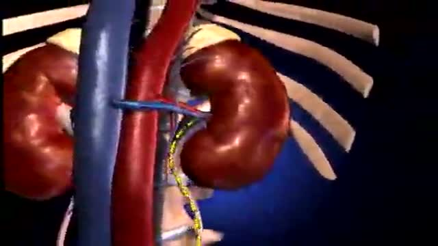
Chronic Renal Failure
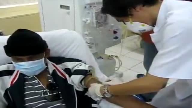
Renal Kidney Hemodialysis
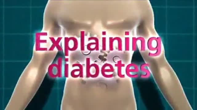
Diabetes Effects on Body Animation 3D
Renal Failure Treatment Options
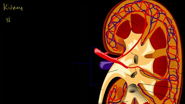
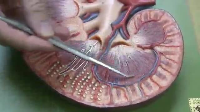
Kidney and Nephron
A great video showing dunctions and physiology of the Nephron
The Nephron Functional Unit of Kidney
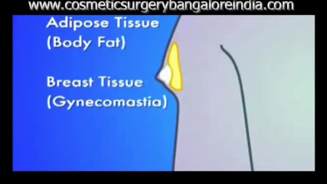
Before and After images following gynecomastia correction with surgical video and animation
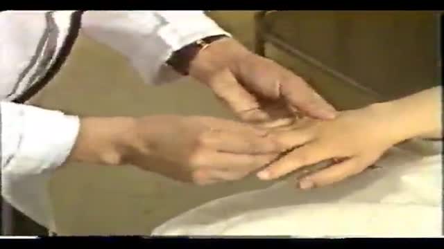
Chinese Complete Physical Clinical Exam
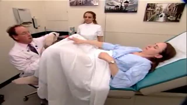
Pelvic Exam Tutorial: Medical Video showing gynecological medical examination of the femal pelvis including bi-manual examintation
Vastibular System Balance

Scientifically Erectile Dysfunction Penile Implants

Diabetic Foot Surgical Debridement

Stop Nose Bleeds by Cautery
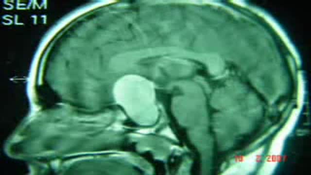
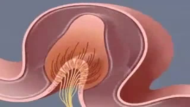
Craniopharyngioma Complete Excision
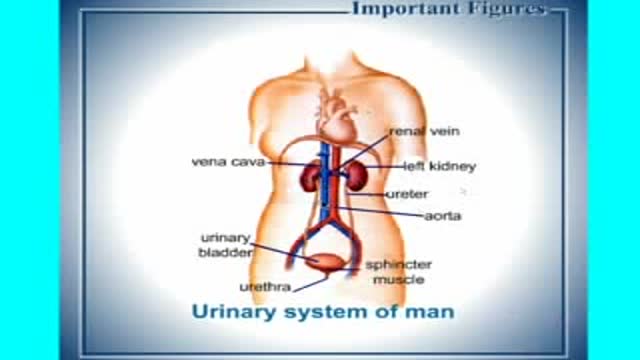
Physiology of Urinary System in Arabic
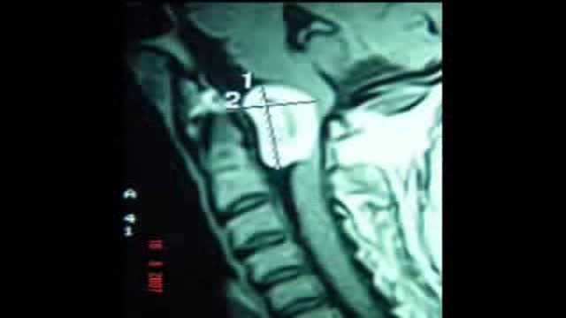
Foramen Magnum Neurofibroma Video
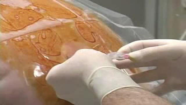
Paramedian Thoracic Epidural Anaesthesia
