סרטונים אחרונים
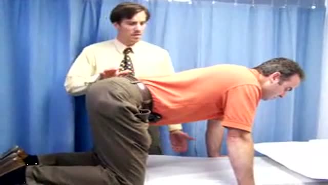
FULL Shoulder Exam by University of Winsconsin
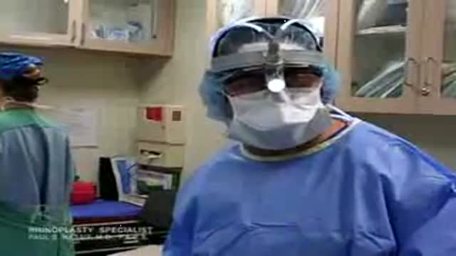
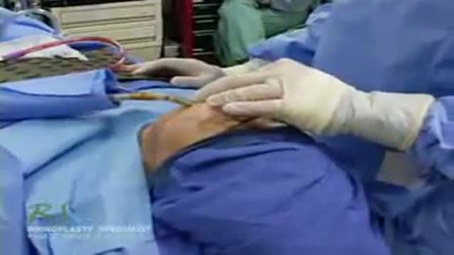
Closed Rhinioplasty Exposing The Nasal Structures
A plastic surgery video showing Turbinal Reduction and Turbinoplasy of the nose
Robotic-assisted endoscopic thyroid surgery using the daVinci® Surgical System can safely and effectively offer those needing thyroid surgery relief without neck incisions. Dr. Ron Kuppersmith and Dr. Andrew deJong are now performing this procedure at the College Station Medical Center in Texas.
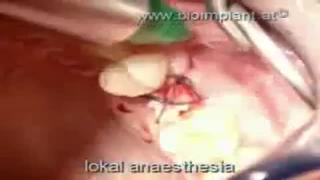
WORLD'S FIRST TRUE ANATOMIC ZIRCONIA DENTAL IMPLANT SOLUTION dentistry
Ultrasound Guided Sclerotherapy for Varicose Veins

Conjoined Twins
Evaluating the Gall Bladder with Ultrasound
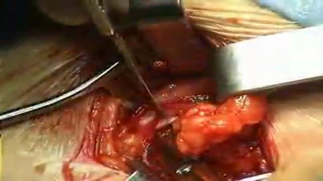
Femoral Hernia Repair with Prosthetic PHS repair placed on anterior way
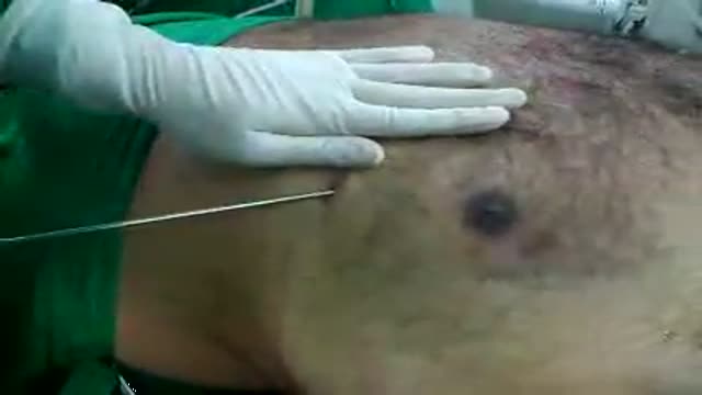
A great video discussing ultrasound guidance of central venous catheter placement
Video demonstrates the action of the isolated lumbar multifidis muscle
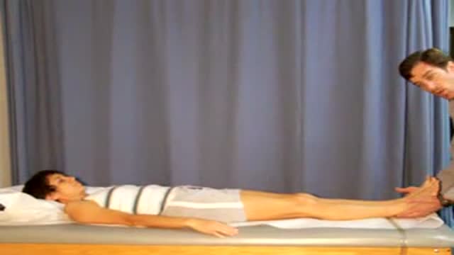
Demonstration of how to differentiate between a true and an apparent leg length difference. The subject is a female with a true short femur.

WORLD'S FIRST TRULY ANATOMIC MULTI-ROOTED ZIRCONIA DENTAL IMPLANT SOLUTION dentistry video
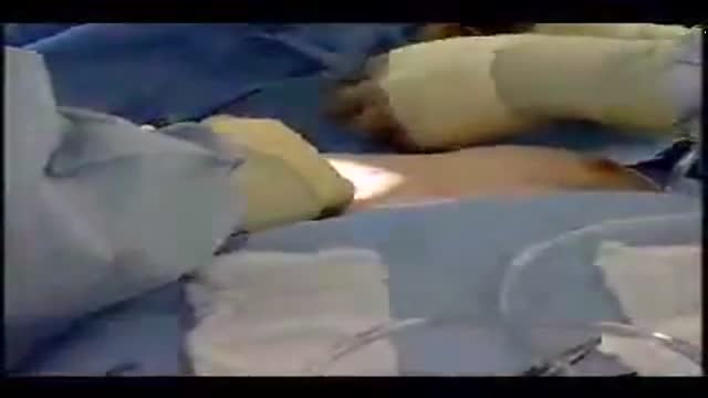
Male Breast Liposuction Reduction
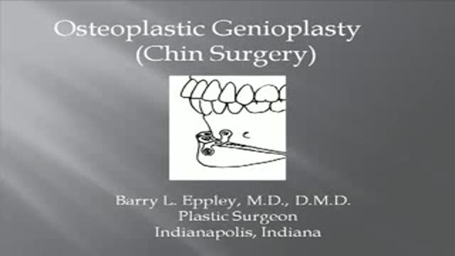
Plastic Surgery of the Chin
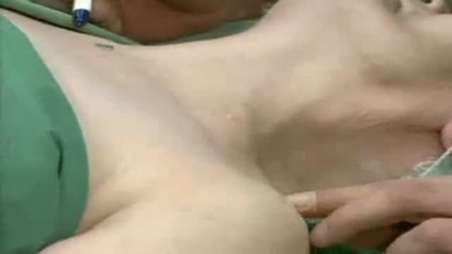
Breast Augmentation Plastic Surgery Video
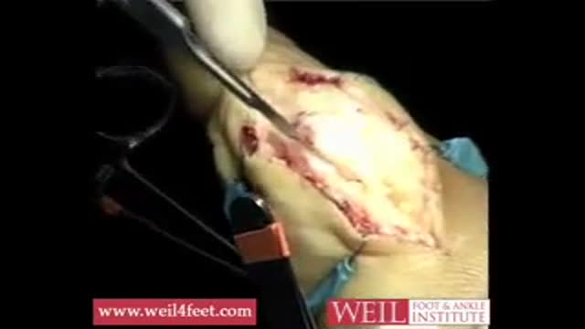
Bunion Correction with Scarf Akin Procedure
A video showing the process of brachial plexus blockage
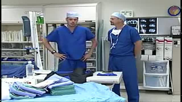
A video showing the accurate steps of Gloving, Gowning and Surgical Scrub
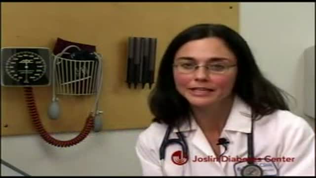
A video discussing the importance of following up the blood pressure for diabetic patients and the serious complications that they can avoid by this very simple measure.
