Latest videos
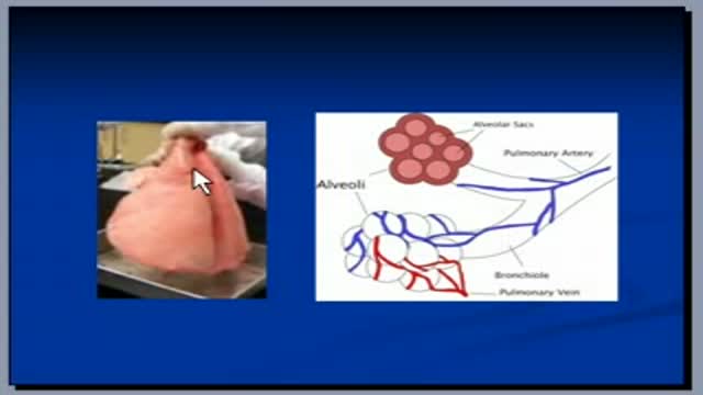
The video will help you to understand radiologic anatomy. Please see my website for disclaimer.
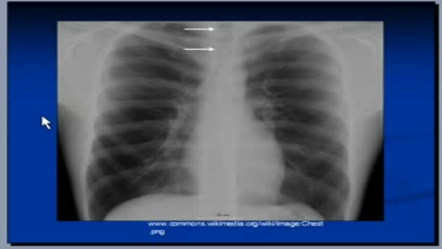
The video will describe mediastinal structures as they are seen on X-ray. Please see web site for disclaimer.

After a bad fall, a patient suffering from spinal fusion seeks help from a DMC Neurosurgery specialist.
~ Detroit Medical Center

An overview of several complex spinal surgeries performed by the DMC Harper University Hospital Department of Neurosurgery. ~ Detroit Medical Center
A teenager's ability to perform the most basic tasks is threatened by a disorder that requires the replacement of both hip joints by a DMC pediatric orthopedic specialist. ~ Detroit Medical Center
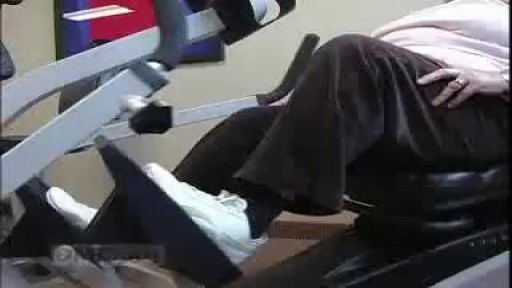
DMC Orthopaedic Specialists are the state leaders in a unique new procedure to resurface the knee joint, preserving more bone for the patient. ~ Detroit Medical Center

Hip Resurfacing Video

Hip Resurfacing Surgery Videos Welcome to the website of the Asian Regional Center for Hip Resurfacing (ARCH) is a specialized surgical center in Apollo Speciality Hospital Chennai. More than 1350 Hip Resurfacing Surgeries have been performed so far. Asian Regional Center for Hip Resurfacing is the first specialized resurfacing center in Asia. Patients with arthritis and hip pain travel from all over the world travel to ARCH for hip surgery. Hip Resurfacing Surgery has revolutionized hip arthroplasty especially for younger and active patients. Unlike conventional Total Hip Replacement (THR) the hip resurfacing conserves the bone in the hip which would be crucial in younger patients. No restrictions are imposed on the resurfaced hip and the patient can participate in any professional or recreational activity after the surgery.

A video show phlebotomy tips
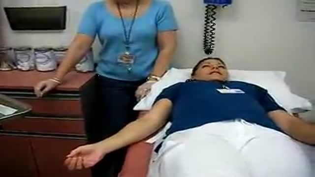
Draw Blood Samples
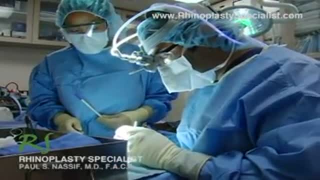
Join Rhinoplasty Specialist Dr. Paul Nassif, a world-renowned expert in revision rhinoplasty, in the operating room as he performs a Columella Strut Placement. His practice, Spalding Drive Cosmetic Surgery & Dermatology, is located in Beverly Hills, CA.
Video Produced by SPORE Medical
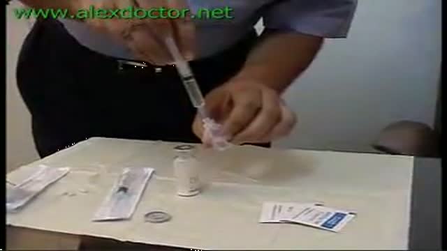
Preparing the Syringe for Injection
Introducing an IM Injection
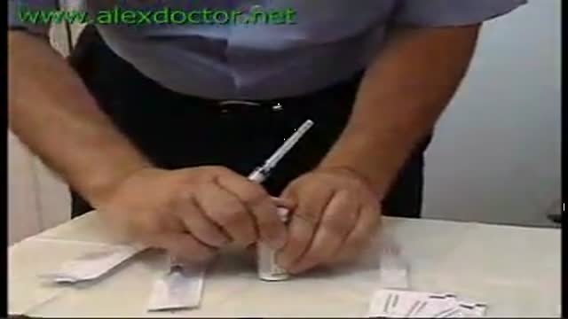
Preparing Syringes for Injections
Brain Stem Tumor Operation
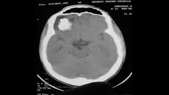
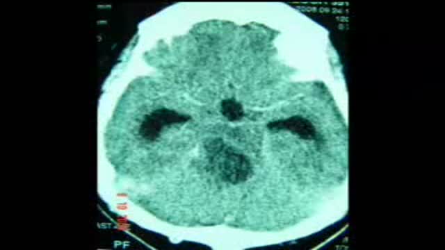
Calcified Brain Abcess complete removal,
surgery
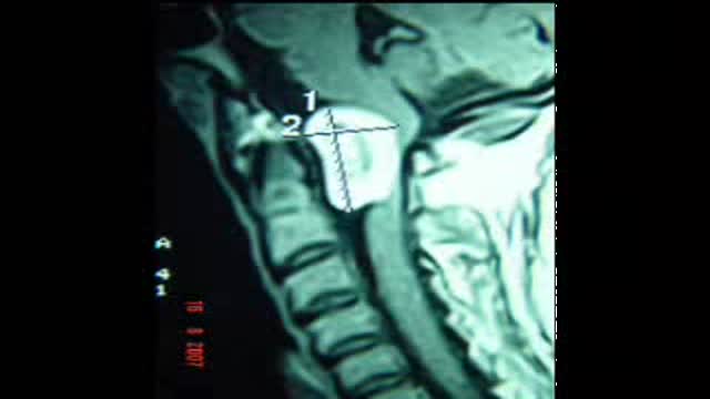
Foramen Magnum Neurofibroma Complete surgical removal.No Deficit
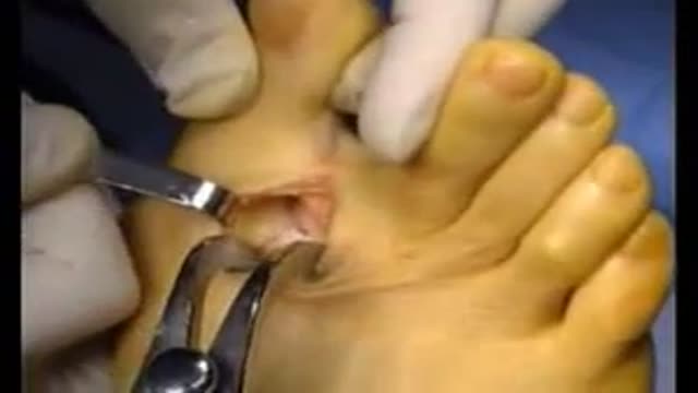
Hallux Valgus Pedis surgery
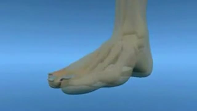
A "Hallux Valgus" or "Hallux Abducto-Valgus" deformity, is commonly referred to as a "Bunion." This describes a pathological condition involving the position of the "hallux" in relation to the first metatarsal.
A bunion deformity can clinically present with a variety of characteristics. The foot itself may present with a wide splaying of the forefoot and a painful bump on the medial aspect of the first metatarsal phalangeal joint. In addition, the hallux may be abducted from the midline of the body, with a valgus rotation in the frontal plane.
A radiographic analysis of a bunion deformity in the Anterior/Posterior or Dorsal/Plantar view will reveal a variety of pathological components. Most notably so, is the exaggerated inter-metatarsal angle between the first and second metatarsal. This may be accompanied by a displacement of the first metatarsal from its position over the sesamoids, such that the metatarsal demonstrates a medial alignment away from the sesamoids which lie to the lateral side.
In some cases, the proximal articular set angle at the head of the first metatarsal may be off-set. This "PASA" is one of the factors which determines the position of the proximal phalanx on the metatarsal during movement as well as at rest.
Although conservative care may involve shoe modifications, padding, strapping, and custom orthosis; surgical reconstruction may be required to alleviate painful and immobilizing bunion conditions.
Soft tissue components of the bunion deformity are primarily addressed by means of a capsular modification, as well as a tenotomy of the adductor tendon at its insertion on the base of the proximal phalanx. The fibular sesamoid may be repositioned by a release of the surrounding ligaments.
Surgical management of the bone or osseous components of a bunion deformity will commonly include an osteotomy and correction to re-establish a more functional position of the first metatarsal within the forefoot. This capital fragment of bone is held in place with hardware fixation in order to secure a proper alignment during the healing phase, thus allowing the hallux to return to a more functionally useful position in the sagittal plane.
