- Physical Examination
- Surgical Examination
- Ophthalmology
- Clinical Skills
- Orthopedics
- Surgery Videos
- Laparoscopy
- Pediatrics
- Funny Videos
- Cardiothoracic Surgery
- Nursing Videos
- Plastic Surgery
- Otorhinolaryngology
- Histology and Histopathology
- Neurosurgery
- Dermatology
- Pediatric Surgery
- Urology
- Dentistry
- Oncology and Cancers
- Anatomy Videos
- Health and Fitness
- Radiology
- Anaesthesia
- Physical Therapy
- Pharmacology
- Interventional Radiology
- Cardiology
- Endocrinology
- Gynecology
- Emergency Medicine
- Psychiatry and Psychology
- Childbirth Videos
- General Medical Videos
- Nephrology
- Physiology
- Diet and Food Health
- Diabetes Mellitus
- Neurology
- Women Health
- Osteoporosis
- Gastroenterology
- Pulmonology
- Hematology
- Rheumatology
- Toxicology
- Nuclear Medicine
- Infectious Diseases
- Vascular Disease
- Reproductive Health
- Burns and Wound Healing
- Other
Latest videos
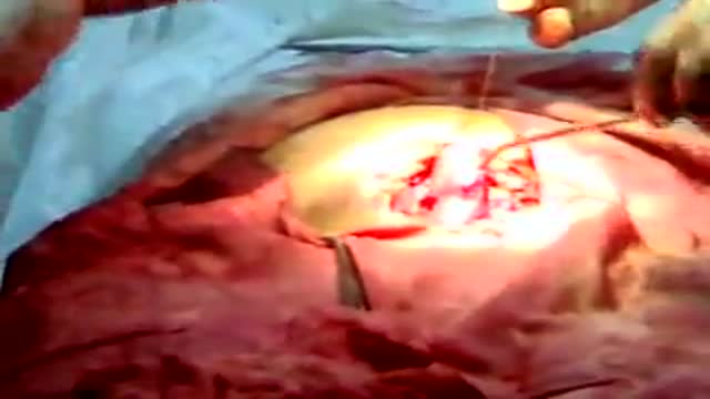
Avideo showing suturing of the uterus and abdominal wall after c-section
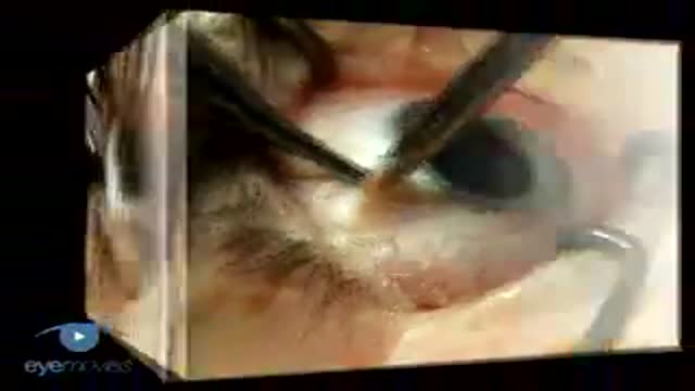
Scleral Buckling: Slinging Muscles & Marking Breaks VR1 Basic Techniques
Off-Pump CABG in Dextrocardia; A New Challenge for a New Era
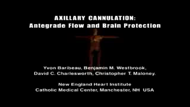
Axillary Cannulation: Antegrade Flow and Brain Protection
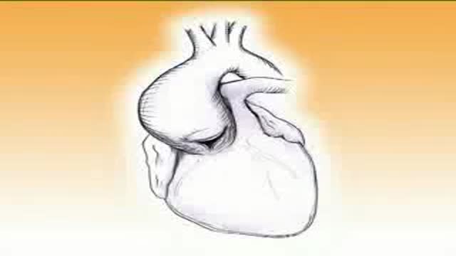
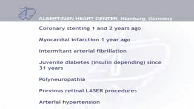
Repair of Anomalous Left Coronary Artery from the Pulmonary Artery (AlCAPA) in an Adult
Aortic Valve-Sparing Operation in a Patient with Aortic Root Aneurysm using a new Prosthesis for Anatomical Reconstruction of the Sinuses of Valsalva
Endoscopic Atraumatic Coronary Artery Bypass EndoACA
Arterial Coronary Off-Pump Revascularization

Sentinel Lymph Node removal in breast Cancer en Français
A video-animation presentation about sentinel lymph node biopsies for breast cancer diagnosis. 3D graphics are used to explain the process. Topics include the lymphatic system and the methods used. This video is part of the breast cancer education series produced by CancerQuest at Emory University
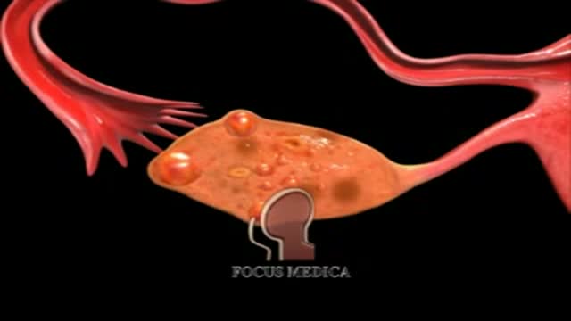
An animation showing what PCO is

Polycystic ovary syndrome (PCOS, also known clinically as Stein-Leventhal syndrome), which is an endocrine disorder that affects 5--10% of women. It occurs amongst all races and nationalities, is the most common hormonal disorder among women of reproductive age, and is a leading cause of infertility. The symptoms and severity of the syndrome vary greatly between women. While the causes are unknown, insulin resistance (often secondary to obesity) is heavily correlated with PCOS.
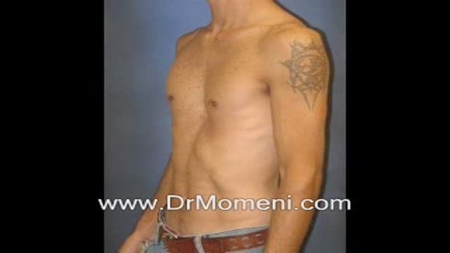
Pectus excavatum (hollow chest) deformity is not uncommon (sometimes mild and other times severe in its form). The chest deformity is often the source of self-consciousness for the patients while growing up. Several surgical techniques (Nuss procedure, Ravitch procedure, etc) are available.
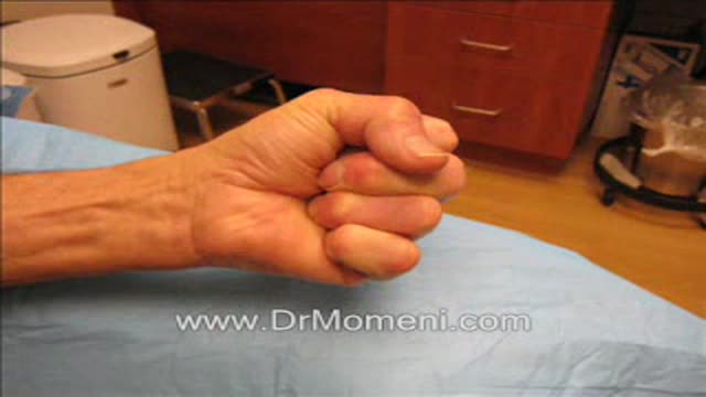
Needle fasciotomy (aponeurotomy) is usually a 15-Minute in-office procedure for Dupuytren's contracture. Performed under local anesthesia, in the office, by board-certified plastic surgeon Reza Momeni, MD. This is a minimally invasive treatment for Dupuytren's.
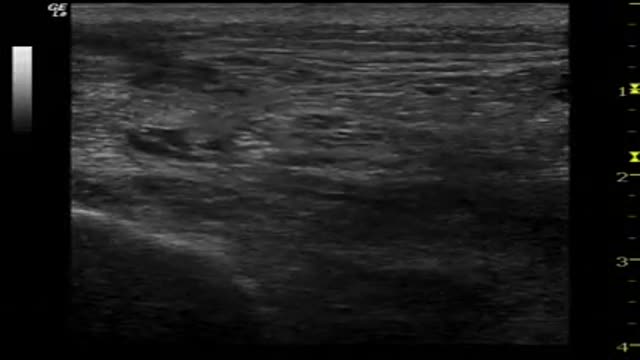
UltraSound-guided Sciatic nerve block by supra popliteal approach
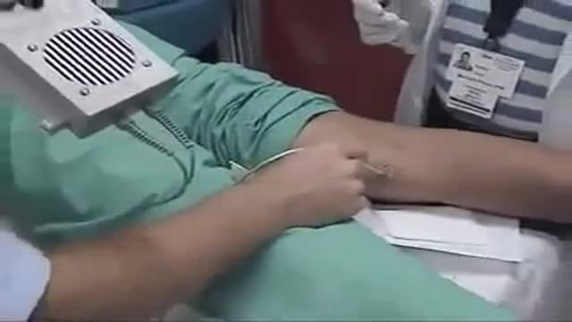
Technique for Popliteal/Peroneal Nerve Block

Ultra Sound-Guided Interscalene Block
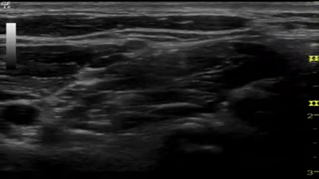
Interscalene Block
Sciatic Nerve Block
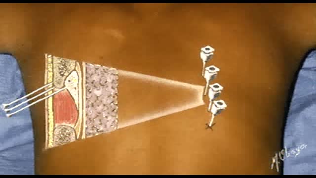
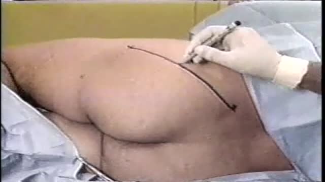
Intercostal Nerve Block
