Senaste videorna
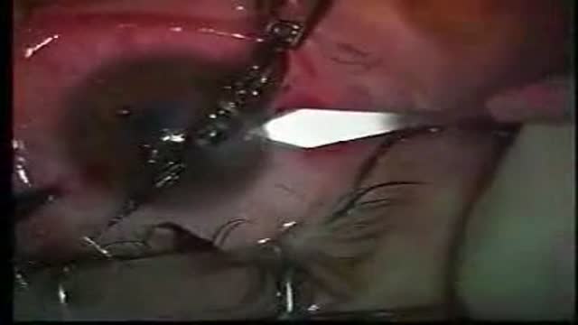
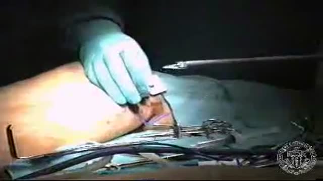
Removal of a foreign body from the eye (fish hook)
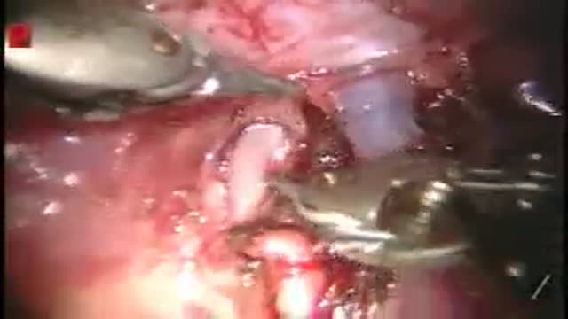
Pulmonary Artery
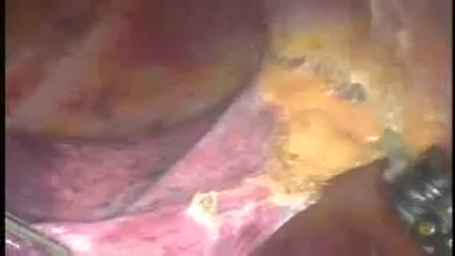
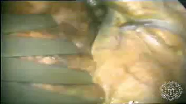
Pulmonary adhesions
Off-Pump Coronary Artery Bypass Grafting (CABG)
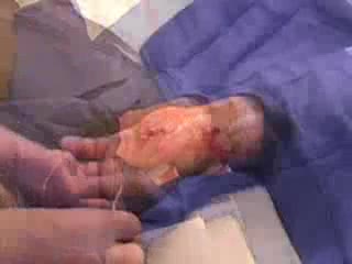
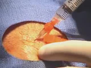
Endoscopic Vein Harvest
Mitral Valve Chorda Repair
Mitral valve repair
Foreign Body(FB) Airway (Whistle) was inhailed by a child causing intermitent stridor & respiratory distress.FForeign Body was removed successfully by rigid endoscopy under General Anesthesia (G/A).The relevant steps of procedure are shown
Video-Assisted thoracoscopy
A video performed by Harvard medical school showing the arterial line placement

A Video from New England Journal of Medicine showing how to do nasogastric intubation
A video showing arthrocentesis of the knee from Harvard medical school
A video showing how to catheter the male urethra
A video from the New England Journal of Medicine performed by Harvard medical school showing Thoracocentesis
A video from the New England Journal of Medicine performed by Harvard Medical School showing basic lacerations repair

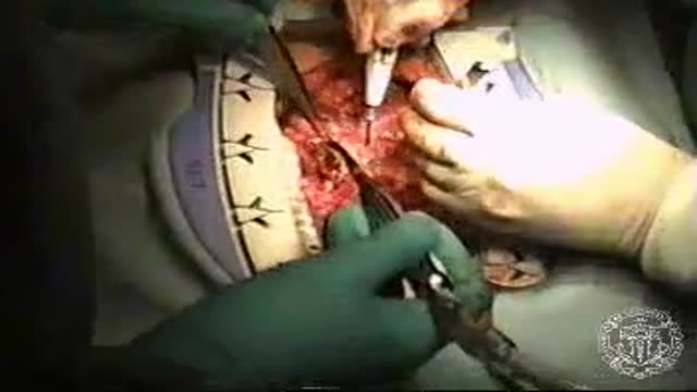
operation on the stomach
a complete discription of the instruments used in laparacopic surgeries and there function
A video from Harvard medical school showing Paracentesis
The video shows how to perform the orotracheal intubation.Performed by harvard medical school
Hip examination by Harvard medical school
