Ultimi video
lowerlimb motor assesment
Examination of the hip
Examination of lumbar spines
Examination of the hand and the wrist
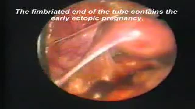
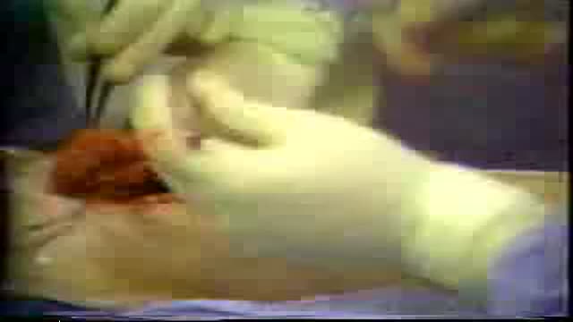
Laparoscopic removal of ectopic pregnancy

full examination of the foot and ankle

Examination of the cervical spines
Examination of peripheral pulses of the lower limb
Complete examination of the abdomen including all the items: inspection, palpation, percussion and auscultation Video
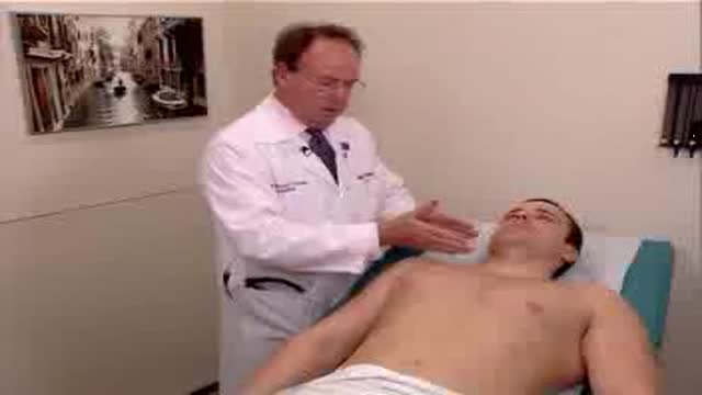
Examination of the heart and lungs with heart sounds
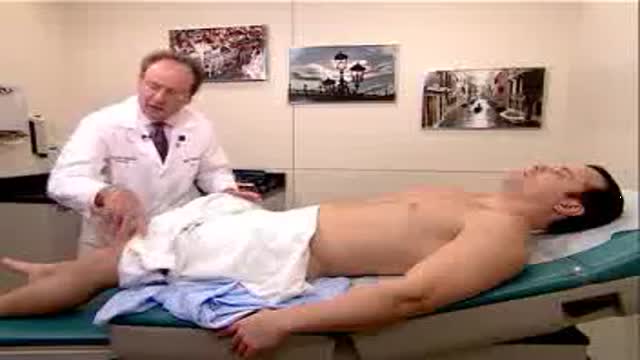
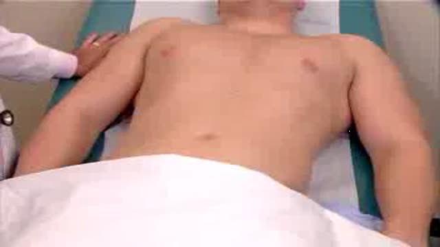
Observation of both jugular veins can provide a reliable indication of the volume and pressure in the right side of the heart since internal jugular veins pulsate in response to phasic changes in right atrial pressure. Proper positioning of the patient to increase the effects of gravity enhances distention of the jugular veins and, therefore, increases the ability to observe venous pulsations.
the most funny medical examination ever..Not: do not try at your office or clinic
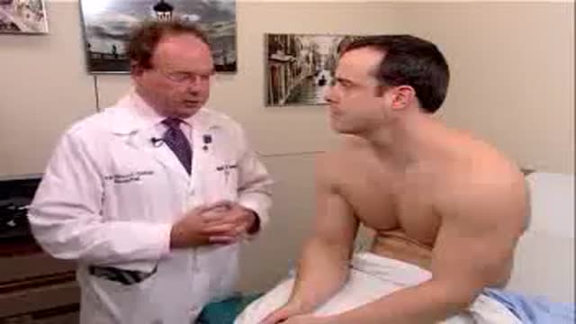
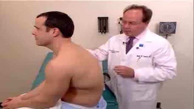
Full complete clinical examination of the chest, lungs and respiration with breath sounds
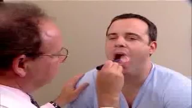
Clinical complete examination of the mouth and throat
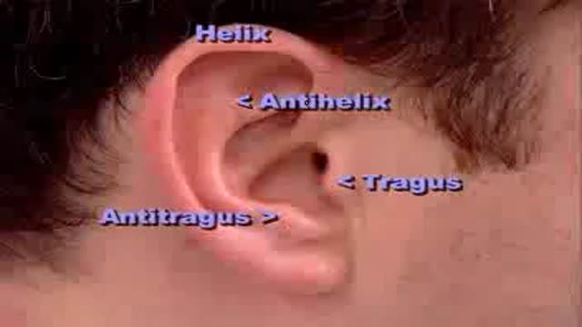
Complete clinical examination of the ears with all the associated tests
Examination of the eye,vision,retina and field of vision

It is very important to instruct your patients about how to self exam their breasts for any abnormalities or masses for early detection of any changes
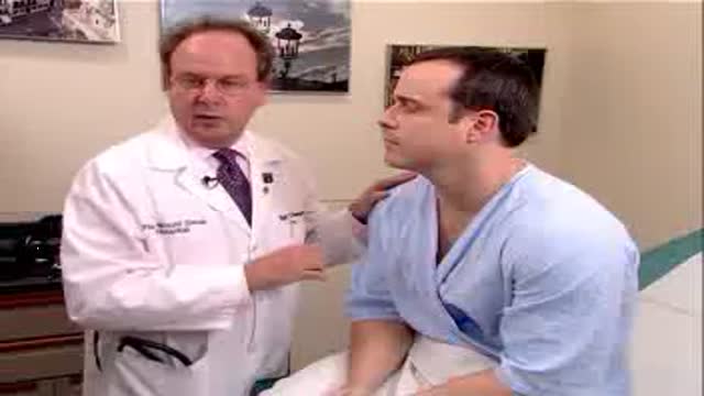
Examination of the lymph nodes of the head
