Latest videos
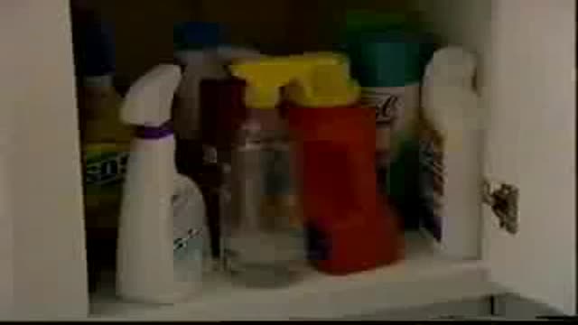
How to deal with a case of ingested poison
Infant Cardio-pulmonary Resuscitation
Infant Airway Obstruction and how to deal with
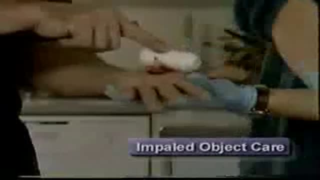
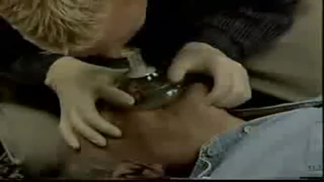
A video showing impaled objects
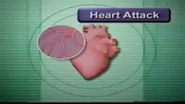
How to deal with heart attack and with stroke
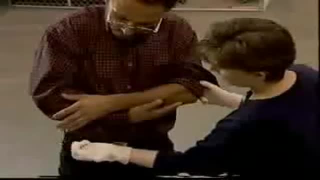
how to to deal with fractures and dislocations

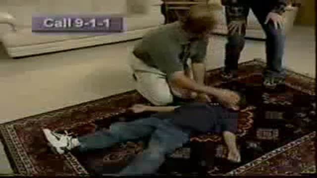
How to move a patient during an accident or during emergency
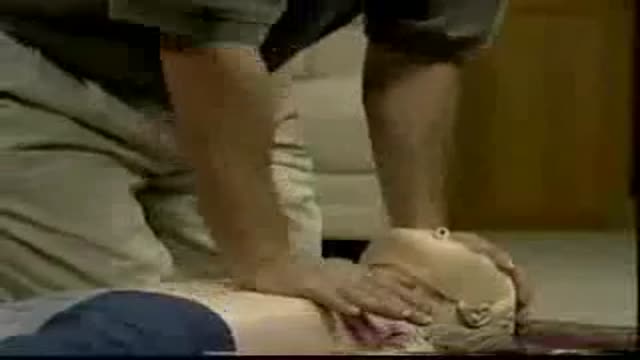
A video showing how to perform Cardio-Pulmonary Resuscitation on a child
Child Unresponsive Airway Obstruction
Child Responsive Airway Obstruction
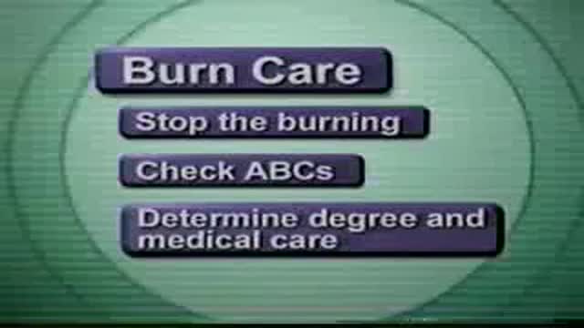
A video shows how to deal with thermal burns
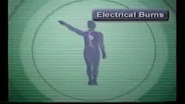
A video showing how to deal with electrical burns and their first aid
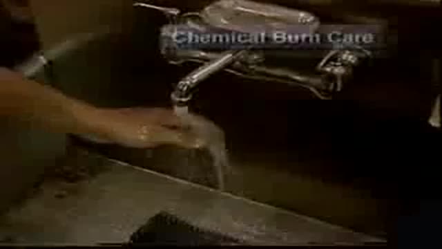
How to deal with chemical burns and their first aid
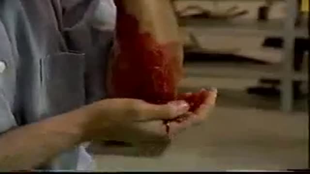
This video shows how to control bleeding

this video is showing the barrier devices
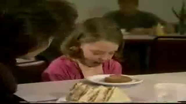
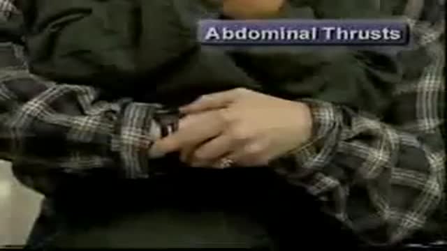
A videos showing Responsive Airway Obstruction and how to deal with that situation
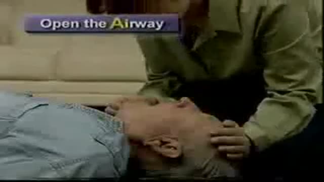
A video showing Unresponsive Airway Obstruction and how to deal with it
http://www.wss4m.com/vb
