- Physical Examination
- Surgical Examination
- Ophthalmology
- Clinical Skills
- Orthopedics
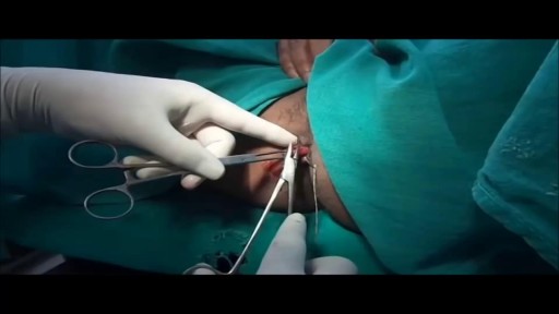
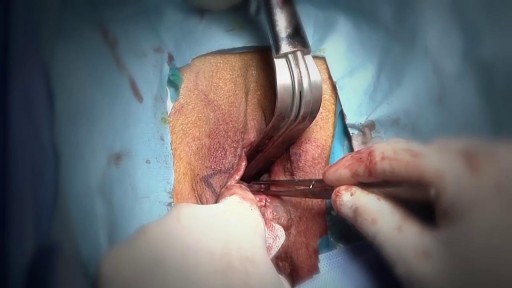
- Surgery Videos
- Laparoscopy
- Pediatrics
- Funny Videos
- Cardiothoracic Surgery
- Nursing Videos
- Plastic Surgery
- Otorhinolaryngology
- Histology and Histopathology
- Neurosurgery
- Dermatology
- Pediatric Surgery
- Urology
- Dentistry
- Oncology and Cancers
- Anatomy Videos
- Health and Fitness
- Radiology
- Anaesthesia
- Physical Therapy
- Pharmacology
- Interventional Radiology
- Cardiology
- Endocrinology
- Gynecology
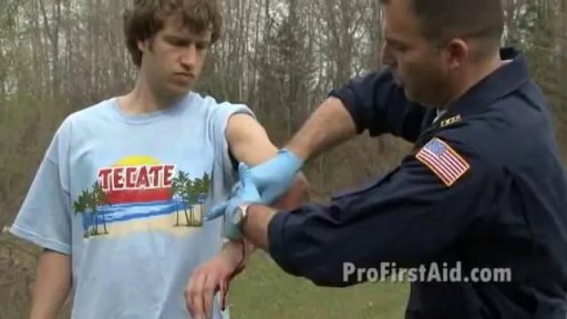
- Emergency Medicine
- Psychiatry and Psychology
- Childbirth Videos
- General Medical Videos
- Nephrology
- Physiology
- Diet and Food Health
- Diabetes Mellitus
- Neurology
- Women Health
- Osteoporosis
- Gastroenterology
- Pulmonology
- Hematology
- Rheumatology
- Toxicology
- Nuclear Medicine
- Infectious Diseases
- Vascular Disease
- Reproductive Health
- Burns and Wound Healing
- Other
Latest videos
Watch that Above Knee Medical Amputation Surgery

Watch that video to know How to Get Pregnant with Twins

Iodine For Ringworm, Best Ointment For Ringworm, Where Do You Get Ringworm, How To Treat Ring Worms ---- http://ringworm-cure.plus101.com --- Ringworms, contrary to the common notion, do not come from worms. Tinea, which is the medical term for ringworms, is a fungal infection seen on the skin's surface. Knowing how to cure ringworm is important because ringworms can be highly contagious. It can be contracted from direct contact with the host (person or animal) as well as by other means such as having contact with the host's clothes. Swimming pools can also be a place where ringworms are transmitted from one person to another. How To Cure Ringworm - Understanding Aspects and Options Different means on how to cure ringworm are available and they sometimes vary in accordance with where the ringworm is located (it can appear in areas like the nails, fingers, toes, feet, scalp, stomach, chest, thighs, and scalp), and the particular type of ringworm. • Ringworms found in the scalp are usually treated with an antifungal shampoo to keep the area dry and clean. • Ringworms found in the feet can be treated through the application of ointments. • Oral medications can also be taken in especially when ringworms are on the nails. • Sprays, powders and creams are also forms by which anti-fungal drugs are bought. These medicines may take some time to work. The infection may persist for a few weeks to several months, depending on the severity and how the body responds to the medications. How To Cure Ringworm - OTC and Prescription Medications Ringworm appears on the skin's surface as an itchy, red, circular patch. As it progresses, it expands and smaller round patches can develop. It is important to immediately identify ringworms and know how to treat them properly. There are many over the counter topical creams (anti-fungal ones) and ointment that can be bought in the market. However, some people prefer to visit the doctor and ask for a prescription. Stronger formulations are generally available via prescriptions. William Oliver is a nutritionist, medical researcher, and author of the Fast Ringworm Cure e-book. To find out how to cure Ringworm in 3 days or less, click below: http://ringworm-cure.plus101.com
Ringworm On Chin, Best Way To Treat Ringworm, How Do I Get Rid Of Ringworm, How To Stop Ringworm --- http://ringworm-cure.plus101.com ---- I'm going to share with you how to cure Ringworm in less than 3 days by using these proven methods that have worked for thousands of people around the world with Ringworm. Not only that, but these same methods work for children, as well as adults, and even pets. Whether you have athlete's foot or jock it, this will work for you, as those are both types of Ringworm. This fungal infection can be cured without drugs or medications. It's important to understand that. In fact, there are many home remedies for Ringworm that have worked for thousands of years for treating many ailments and skin conditions. Below I will list a few. First, understand there are 3 primary methods to cure Ringworm. You must do all 3 if you want to know how to cure Ringworm as fast as possible. Over the 5 years I've spent studying and learning about Ringworm, I've tried almost every treatment and home remedy available. I've found this to be the most effective and fastest way to cure Ringworm. 1) Treat the rash with home remedies and treatments. There are many home remedies for Ringworm available, as well as natural treatments, that are good for the skin and can promote healing rapidly. Not only do they get rid of the itchiness, pain or discomfort, but they also heal the rash and prevent it from coming back again. 2) Use bathing procedures. Bathing procedures are a powerful way to cure Ringworm, especially when you add home remedies and ingredients to the bath. This again, promotes healing, and helps get rid of all symptoms associated with Ringworm. 3) Consume the right foods and supplements. A proper diet is important to cure Ringworm. Treating Ringworm externally is not enough - you need to provide the body with what it needs to fight off the infection and boost the immune system, so you never have to worry about it again. Nutrition, hydration, rest, and supplementation are all equally important. Follow these 3 methods and you will be cured from Ringworm within days. If you sit and wait, and do nothing, it will only get worse and you will suffer much longer than you need to. To find out more on how to cure Ringworm in 3 days or less, check out the Fast Ringworm Cure e-book program that goes into all the ways to cure Ringworm fast, along with a list of home remedies and treatments that you can apply right away. William Oliver is a nutritionist, medical researcher, and author of the Fast Ringworm Cure e-book. To find out how to cure Ringworm in 3 days or less, click below: http://ringworm-cure.plus101.com
What Gets Rid Of Ringworm, How To Cure Ringworm On Face, How To Cure Ringworm Naturally, Ringworms ---- http://ringworm-cure.plus101.com --- There are many cures, treatments and home remedies available at your disposal. While most doctors prescribe some type of weird fungus cream or medication, this isn't always the best route and can do more harm to your body than good. Think about it, anything that isn't natural and is made up of chemicals isn't going to nourish and provide your body with the strength it needs. The worst thing you can do is to "sit and wait" for it to magically heal on it's own. When people do this, they're often in for a lot more pain and discomfort than they need to go through. You want this Ringworm condition over and done with quickly, so that you can move on with your life and have this be something of the past. Over the years I've spent studying and researching Ringworm, I've tested almost every treatment and remedy available on the market. I began exploring home-made remedies, as they are more ideal for your body, are inexpensive, and have been around for centuries. You'd be amazed at what our planet provides us and the power these natural ingredients can have in healing our bodies. I've broken down the cure for Ringworm into several steps: 1) Treat the rash immediately by getting rid of any itchiness, discomfort and pain. I suggest bathing procedures for this, which are mentioned in my Fast Ringworm Cure e-book program. A bath, combined with special home remedies and ingredients, can get rid of all symptoms quickly and begin healing the rash rapidly. 2) Use natural creams, lotions, or even oils on the rash. This will help kill the fungus and heal the rash quickly. There are certain ingredients, such as special oils, honey, and many others that have powerful healing affects. 3) Strengthen the immune system and body so that it can naturally fight off any infection and heal the body from the inside out. This is often something overlooked, as most people think of a skin condition as an external thing. But by consuming the right foods, while avoiding others, you can heal Ringworm much faster. Certain supplements that your body may be deprived of are key. Not only that, but a strong immune system and body means that you won't have to worry about getting Ringworm, or any other condition for that matter, again in the future. William Oliver is a nutritionist, medical researcher, and author of the Fast Ringworm Cure e-book. To find out how to cure Ringworm in 3 days or less, click below: http://ringworm-cure.plus101.com
Watch that video to know How To Remove Plaque Without Visiting The Dentist
Watch that video to learn how to stop arterial bleeding
Watch that Hemorrhoids Repairing Medical Video
Watch that Female Genital Walls Tightening Plastic Surgery

Watch that video of a Man Impaled by Shovel in His Butt Inside ER

Watch that video of a Woman Was Pregnant For 46 Years

Watch that video of Terrible Things Were Found Living Inside a Human Body
Watch that Poisoned Human Body Medical Dissect
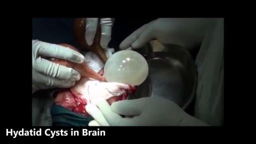
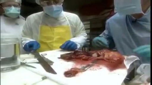
Watch that video of the Worst Brain & Liver Cysts Removal\
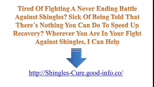
Is Shingles Contagious, What Are Shingles, Herpes Zoster Pictures, Shingles Home Remedies --- http://shingles-cure.good-info.co/ --- If You Are A Newcomer To This Disease, I Hate To Be The Bringer Of Bad News But You Should Know That The List Of Potential Symptoms Is Depressingly Long. These Include The Following: A General Feeling Of Muscle Pain To Begin With, Almost Like Flu A Tingling, Burning Type Sensation In A Specific Area Of The Skin Fever And Headache And Sometimes A Swelling Of The Lymph Nodes A Band Of Spots And Then A Rash At A Specific Part Of Your Body – Very Often The Head Or The Side Of The Trunk Infection Over The Site Of The Rash – Leaving It Prone To Additional Tissue Damage From Bacteria Postherpetic neuralgia leading to chronic nerve pain Ulceration Of The Eye – In Those Cases Where The Shingles Rash Occurs In The Area Of The Eye – Known As Zoster Ophthalmicus. Stress And Depression – Particularly Where The Illness Lingers On For A Long Period Everyone Is Different And Not Everyone Will Experience All Of Those Symptoms. However Even The Most Mild Case Of Shingles Can Be Extremely Debilitating – Something Of Which I Am All Too Aware. Tired Of Fighting A Never Ending Battle Against Shingles? Sick Of Being Told That There´s Nothing You Can Do To Speed Up Recovery? Wherever You Are In Your Fight Against Shingles, I Can Help In this presentation, shows you some unique and rare methods to get rid of shingles naturally in as little as 14 days! This is based on proven techniques used by shingles sufferers without the use of pills and other medication. Get Rid of Shingles will also boost your energy and health dramatically and improve the quality of your life. IMPORTANT NOTE: I can't leave this video up for long, so be sure to watch it from beginning to end while it's still here. REMEMBER: Watch the whole video, as the ending will pleasantly surprise you. click here: http://shingles-cure.good-info.co/

Pictures Of Shingles, Images Of Shingles, Cause Of Shingles, Can You Catch Shingles, Cause Shingles --- http://shingles-cure.good-info.co/ ---- Home Remedy For Shingles Treatment. There are surprisingly diverse amounts of treatments that can be used for viruses, especially one in particular known as Shingles. Shingles is the type of virus that is annoyingly unpredictable. Some people get it while others do not, and while it predominant after a certain age, most people can go their whole lives without ever seeing a hint of it. Yet, we all have it living inside of us. The first step to treating the virus correctly is to understand what can set it off. The first pre-requisite is to have had chicken-pox before. Most things manage to squeeze through an immune system if it is weak enough and the shingles virus is no different. In some cases that can kill someone with a weak enough system The shingles virus can also ‘wake up’ if sufficient levels of stress agitate a person’s immune system. As mentioned before, the virus is unpredictable, and even if you meet all the requirements you could go through your entire life without one outbreak. The second step to treating the virus is catching it in time. 72 hours after you first begin to notice symptoms of shingles is pushing your luck so within that time frame some type of treatment should be instigated. For the most part, for a disease like shingles, the main source of relief comes from skin creams and pain killers. Neither of which is a cure for the pain, but which do happen to fall under the heading of ‘home remedy’ Wet rags can be used to soothe the inflamed and tender skin, and a binding with a substance known as aluminum acetate can protect the infected area without causing further irritation. Calamine lotion is a fail safe for almost any sort of skin irritation so it would be smart to go through the aisles at your local grocery store to find products similar. Then of course there are the most obvious measures that need to be taken during your do it yourself treatment sessions. Stay out of the sun since the heat and UV rays can cause unbearable pain against already brutalized skin. Also, keep yourself from scratching at the area since, like the chicken-pox, you will only end up making it worse. If you do not believe me then consider the unlucky individuals who do not have the rash on a small area, but rather all over their bodies. Anti-itch cream would probably be more than appreciated at that point. Should the time come where the virus is making repeated appearances, then it may be time to throw in the towel and head for the hospital. The reason is that after the first outbreak, it isn’t unusual that the virus that was originally stored in the roots of your nerves ‘burned’ itself out. However, continual outbreaks are indications of something deeper that can only grow worse as the attacks continue. Some victims of the virus have described the sensation as having your flesh eaten from the inside out. Once the pain reaches that level, stubbornness should be put on the backburner. If you are forced to go to your local doctor, you may be provided with some of the medicines that have recently been created to help fight off shingles. They are by no means brand names, but neither are they over the counter drugs either. After a few more years to study their effectiveness, there is no reason why this new medication cannot ease the pain caused by shingles, and even, eventually cure it. In this presentation, shows you some unique and rare methods to get rid of shingles naturally in as little as 14 days! This is based on proven techniques used by shingles sufferers without the use of pills and other medication. Get Rid of Shingles will also boost your energy and health dramatically and improve the quality of your life. IMPORTANT NOTE: I can't leave this video up for long, so be sure to watch it from beginning to end while it's still here. REMEMBER: Watch the whole video, as the ending will pleasantly surprise you. click here: http://shingles-cure.good-info.co/
Watch that video to know Medical Hazards and Risks of Anal Intercourse

Generalized Anxiety Disorder, Symptoms Of Anxiety Attack, Shortness Of Breath Anxiety --- http://panic-attacks-anxiety.good-info.co --- Newly Discovered Panic "Off Switch" Gives You Anxiety Relief Without Pills or Therapy Here's an interesting fact about anxiety and panic attacks: Did you know that just like the hiccups, doctors still can't agree exactly why they happen to you? And did you also know there's a 60-second solution to panic and anxiety that you can do anywhere? Yes, it takes you just one minute and I'm going to share it with you today. Until one day about a year ago, I thought I might be doomed to let panic attacks rule my life. And I made this free online presentation to tell you about the one discovery about panic and general anxiety that finally cut through the confusion and changed everything. Pay very close attention, because whether you've only had one or two "attacks" so far… or even if you've been having them for years and it seems like a life sentence you'll never escape from… You're about to discover one weird thing that panic, anxiety and the hiccups – yes, the hiccups – have in common that goes right back to the stone age. Discover How To Begin Eliminating Panic And Anxiety From Your Life Forever Click Here: http://panic-attacks-anxiety.good-info.co

Symptoms Of Anxiety, Anxiety Disorder Symptoms, What Is Social Anxiety, Zoloft For Anxiety --- http://panic-attacks-anxiety.good-info.co --- Panic attacks and anxiety While there are times for doctors, I want you to consider this: MOST of your anxiety is under the radar… Masquerading as “just feeling a bit nervous”... or “just a tad irritable thanks to this diet plan”... or, “let’s skip the party and just stay home tonight.” And, I’m telling you, that anxiety not only destroys your fat burning power: It often leads to all-out panic disorder, if you just ignore it. Fortunately for you, there’s a 60-Second Solution that restores your calm, removes those anxious feelings, and allows you to keep burning body fat for energy. Now, this exact same technique works for all-out panic and even more serious anxiety issues, too… The man who delivers this presentation had panic attacks in “everyday” situations… and he too had feelings of anxiety whenever he tried to diet-off body fat… This Simple Trick Stops Panic Attacks And Anxiety Click Here: http://panic-attacks-anxiety.good-info.co
