- Physical Examination
- Surgical Examination
- Ophthalmology
- Clinical Skills
- Orthopedics
- Surgery Videos
- Laparoscopy
- Pediatrics
- Funny Videos
- Cardiothoracic Surgery
- Nursing Videos
- Plastic Surgery
- Otorhinolaryngology
- Histology and Histopathology
- Neurosurgery
- Dermatology
- Pediatric Surgery
- Urology
- Dentistry
- Oncology and Cancers
- Anatomy Videos
- Health and Fitness
- Radiology
- Anaesthesia
- Physical Therapy
- Pharmacology
- Interventional Radiology
- Cardiology
- Endocrinology
- Gynecology
- Emergency Medicine
- Psychiatry and Psychology
- Childbirth Videos
- General Medical Videos
- Nephrology
- Physiology
- Diet and Food Health
- Diabetes Mellitus
- Neurology
- Women Health
- Osteoporosis
- Gastroenterology
- Pulmonology
- Hematology
- Rheumatology
- Toxicology
- Nuclear Medicine
- Infectious Diseases
- Vascular Disease
- Reproductive Health
- Burns and Wound Healing
- Other
Top videos
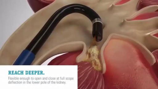
Kidney Stone Breaking Device video, very interesting
Splenectomy for massive splenomegaly (>1500 g) provides palliation but is associated with a high rate of perioperative complications in a population of patients with advanced hematological malignancies. Predictive factors for survival and whether the palliative goals are achieved in the long-term are not well defined.

-Failure to thrive (FTT) is not a diagnosis in itself; rather, it is a term used to describe failure to gain weight in children younger than two years old. Children categorized as FTT weigh less than the 5th percentile for their age; more severe cases involve a slowing of linear growth and head circumference as well. The three causes of FTT are inadequate calorie intake, inadequate calorie absorption, and increased calorie requirements. Newborn infants need 110 kcal/kg/day, while children up to twelve months need 100
Hernia Repair with Prolene Hernia System

Amniocentesis,before the actual procedure, a local anesthetic is sometimes given to relieve the pain when inserting the needle used to withdraw the fluid. A needle is usually inserted through the mother's abdominal wall or at the end of the vagina, and through the wall of the uterus into the amniotic sac. With assistance from ultrasound, a physician aims towards an area of the sac that is away from the fetus and extracts a small amount of amniotic fluid for testing. The puncture heals, and the amniotic sac replenishes the liquid over a day or so. After the amniotic fluid is extracted, the fetal cells are separated from it using a centrifuge, and the fetal chromosomes are examined for abnormalities. Various genetic testing may be performed, but the three most common abnormalities tested for are Down's syndrome, Trisomy 18 and spina bifida. Amniocentesis can be performed as soon as sufficient amniotic fluid surrounds the fetus to allow a sample to be recovered relatively safely, usually no earlier than the 14th week of pregnancy. Often, genetic counseling is offered in conjunction with amniocentesis.
Sutureless laparoscopic radical Prostatectomy

Shave Your Pubic Hair
Definition. The principal signs of cerebellar dysfunction are the following: Ataxia: unsteadiness or incoordination of limbs, posture, and gait. A disorder of the control of force and timing of movements leading to abnormalities of speed, range, rhythm, starting, and stopping.
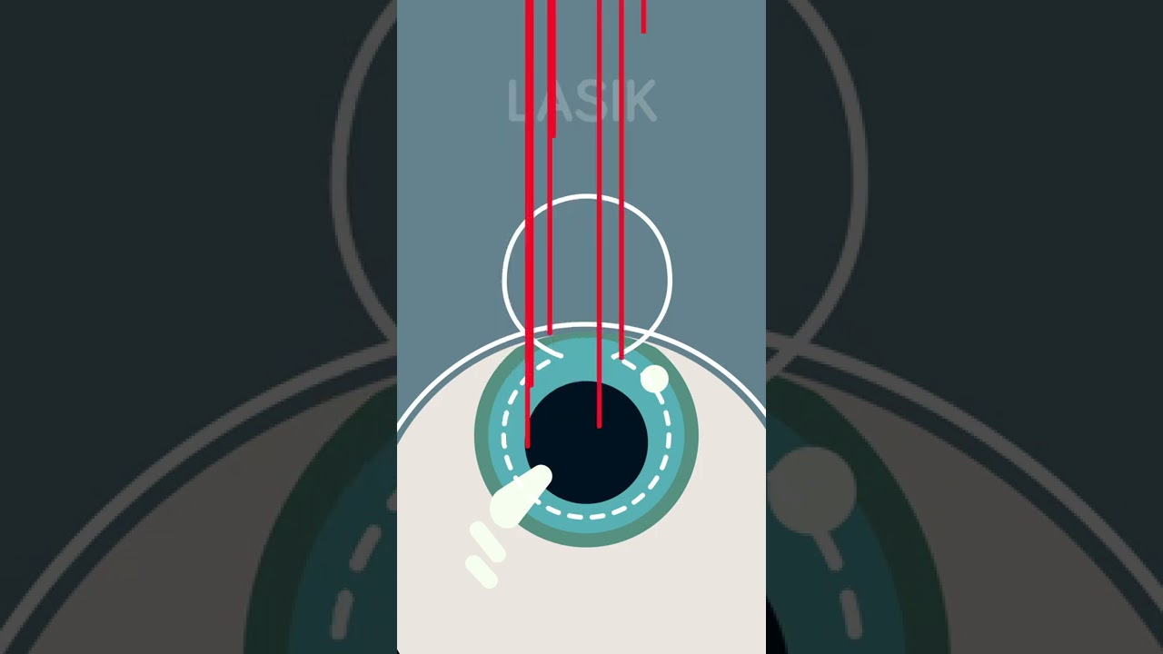
Ever considered getting laser eye surgery, but didn’t know how it worked? Allow us to help!
There are three different main types of laser eye surgery: LASIK, SMILE, and Surface Laser Treatments, and each can be explained pretty easily.
LASIK uses two lasers to open up a thin flap on the surface of the cornea, and then reshapes the cornea underneath. The flap is then placed back over the reshaped cornea, and heals independently with time.
SMILE uses one laser to reshape the cornea through a small, self-healing hole.
And Surface Eye Treatments remove the clear skin over the eye, to then reshape the cornea underneath with - you guessed it - a laser!
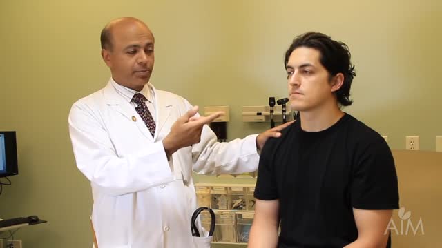
The Knee Exam
Observation:
1. Make sure that both knees are fully exposed. The patient should be in either a gown or shorts. Rolled up pant legs do not provide good exposure!
2. Watch the patient walk. Do they limp or appear to be in pain? When standing, is there evidence of bowing (varus) or knock-kneed (valgus) deformity? There is a predilection for degenerative joint disease to affect the medical aspect of the knee, a common cause of bowing. Varus Knee Deformity, more marked on the left leg. 3. Make note of any scars or asymmetry. Chronic/progressive damage, as in degenerative joint disease, may lead to abnormal contours and appearance. Is there obvious swelling as would occur in an effusion? Redness suggesting inflammation? 4. Is there evidence of atrophy of the quadriceps, hamstring, or calf muscle groups? Knee problems/pain can limit the use of the affected leg, leading to wasting of the muscles.
While both legs have well developed musculature,
the left calf and hamstring are bulkier than the right. 5. Look at the external anatomy, noting structures above and below the knee itself: 1. Patella 2. Patellar tendon 3. Quadriceps/Hamstring/Calf muscles 4. Medial and lateral joint lines. 5. Femur and Tibia 6. Tibial tuberosity
Ballotment (helpful if the effusion is large) 1. Slightly flex the knee which is to be examined.
2. Place one hand on the supra-pateallar pouch, which is above the patella and communicates with the joint space. Gently push down and towards the patella, forcing any fluid to accumulate in the central part of the joint.
3. Gently push down on the patella with your thumb.
4. If there is a sizable effusion, the patella will feel as if it's floating and "bounce" back up when pushed down.
HYSTERECTOMY RECOVERY: ALL PROCEDURES ARE NOT CREATED EQUAL Too often, women are only given the option of an open hysterectomy for conditions like large fibroids or an enlarged uterus. Surgical techniques have evolved in the last decade, but across the United States, the number of women still having open hysterectomy procedures is unnecessarily staggering. Robotic procedures are becoming more common as hospitals invest nearly $2 million in the machine. While the robot does allow surgeons who are not necessarily trained in laparoscopic procedures to perform a more minimally invasive surgery, tools cannot replace skill. There is no added benefit to the patient and the surgery can cost on average up to $2,000 more than other laparoscopic options, and in some cases much higher.
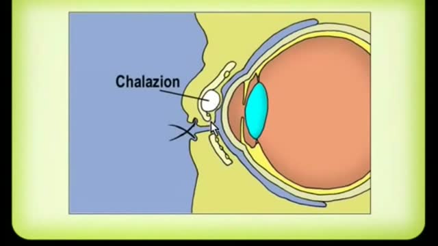
A chalazion is a lump of the lid that is caused by obstruction of the drainage duct of an oil gland within the upper or lower eyelid. This lump may increase in size over days to weeks and may occasionally become red, warm, or painful.

Children are special patients, and their medical needs are unique, including their surgical needs. At UNC Hospitals, an expert and experienced team of physicians treat children in a kid-friendly and family-centered environment. UNC Pediatric Surgeon Dr. Timothy Weiner explains
Understand how this world-class surgery platform operates a minimally invasive robotic surgery during a medical procedure for prostate cancer.
Of the many factors that affect your compatibility with a man, one of the biggest (or smallest) is in his pants. As with humour, interests or habits, the wrong fit can leave you cold. Or traumatised. In a study of 1,661 penises, Dr Debby Herbenick, author of Sex Made Easy, found an almost nine-inch difference in erection size: from 1.6 inches to 10.2. And since absolutely nothing outside the package tells you what to expect with the package, you have to test compatibility the hard way. Sometimes you hit your jackpot, sometimes it's just fine, and sometimes he's the guy on either end of that erection spectrum. These writers have been there, so here's what they learned - and how you can deal (without the gasp reflex).

Common causes of the knee pain
Knee pain is very common and in this video we will present the most common problems that can cause pain in the knee. (Patella) itself, which is in front of the knee, or from the tendons that are attached to the kneecap (patellar tendon and quadricep tendon). One of the most common problems is patellar chondromalacia which is chronic pain due to the softening of the cartilage beneath the kneecap. The cartilage of the kneecap will have some erosions, defects, or holes from mild to complete inside the joint (exactly in the back of the kneecap).
• Pain in the front of the knee
• Occurs more in young people
• Becomes worse from climbing up stairs and going downstairs
Treatment is usually nonsteroidal anti-inflammatory medication, physical therapy, and surgery is very rare. Also in front of the kneecap, the patient may get pain due to prepatellar bursitis.
When there is prepatellar bursitis, the patient will see that the swelling, the inflammation, and the pain is located over the front of the kneecap. The bursa becomes inflamed and fills with fluid at the top of the knee, causing pain, swelling, tenderness and a lump in that area on top of the kneecap. If the pain is in front of the knee but below or above the patella, this may indicate that the patient has tendonitis. Patellar tendonitis is an overuse condition that often occurs in athletes who perform repetitive jumping activities. Patellar tendonitis is a knee pain that is associated with focal patellar tendon tenderness and it is usually activity related. It is located below the kneecap and is called "jumper's knee". Patellar tendonitis affects approximately 20% of jumping athletes. There will be tenderness to palpation at the distal pole of the patella in extension and not in flexion. Quadriceps inflexibility, atrophy and hamstring tightness are predisposing factors for this condition. Treatment is rest, anti-inflammatory medication, stretching and strengthening of the hamstrings and quadriceps. Use an eccentric exercise program. The early stages of patellar tendonitis will respond well to nonoperative treatment. Another important cause of knee pain is a meniscal tear. The meniscus is the cushion that protects the cartilage in the knee. Injury will cause pain on the medial or the lateral side of the knee exactly at the level of the joint. The patient will complain of a history of locking, instability and swelling of the knee. McMurray test will be positive. A painful pop or click is obtained as the knee is brought from flexion to extension with either internal or external rotation of the knee. Arthritis of the knee Knee arthritis is very common. The cartilage cells die with age and its repair response decreases in the joint collapses with increased breakdown of the framework of the cartilage. The patient will have progressive blurring away of the cartilage of the joint with decreased joint space as seen on x-rays. Another source of pain is the Baker's cyst. The cyst is in the back of the knee between the semimembranosus yes and the medial gastrocnemius muscles. Another important source of knee pain is a ligament injury. Here is a normal knee without a ligament injury. Here you can see from the front, you can see the lateral and medial collateral ligament. You can see the ACL and PCL from the side view. These ligaments are usually injured as a result of a sports activity. Here is an example of a sports knee injury. Here is an example of the medial collateral ligament injury. This is the most commonly injury knee ligament injury to this ligament is on the inner part of the knee. Here is an example of an injury of the anterior cruciate ligament. It involves a valgus stress to the knee. Lachman test is usually positive, and MRI is diagnostic. Another important cause of knee pain is iliotibial band syndrome of the knee. Inflammation of the thickening of the iliotibial band results from excessive friction as the iliotibial band slides over the lateral femoral condyle. The iliotibial band is a thick band of fascia that extends along the lateral thigh from the iliac crest to the knee. And as the knee moves, the IT band was repeatedly shifted forwards and backwards across the lateral femoral condyle. The patient will complain of swelling, tenderness, and crepitus over the lateral femoral condyle. The condition occurs in the ITB S occurs in runners, cyclist and athletes that require repeated knee flexion and extension. The pain may be reproduced by doing a single-leg squat. The Ober's test is used to at assess tightness of the iliotibial band. MRI may show edema in the area of the ITB. Treatment is usually nonoperative with rest and ice, physical therapy, with stretching, proprioception, and improvement in neuromuscular coordination. Training modification and injections may be helpful. Surgery is a last resort. Surgical excision of the scarred inflamed part of the iliotibial band.
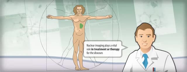
Nuclear medicine is a branch of medical imaging that uses small amounts of radioactive material to diagnose and determine the severity of or treat a variety of diseases, including many types of cancers, heart disease, gastrointestinal, endocrine, neurological disorders and other abnormalities within the body.

Function. Vitamin A helps form and maintain healthy skin, teeth, skeletal and soft tissue, mucus membranes, and skin. It is also known as retinol because it produces the pigments in the retina of the eye. Vitamin A promotes good vision, especially in low light. Vitamin deficiency anemia occurs when your body doesn't have enough of the vitamins needed to produce adequate numbers of healthy red blood cells. Red blood cells carry oxygen from your lungs throughout your body. If your diet is lacking in certain vitamins, vitamin deficiency anemia can develop.
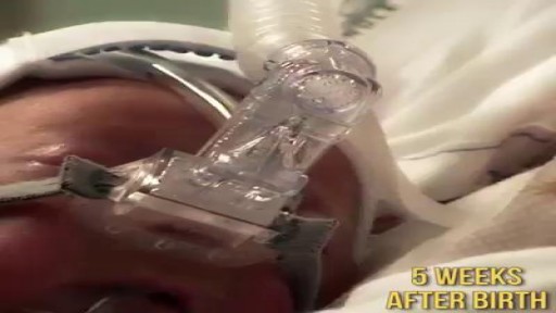
Meet Toby, the baby who was born premature at 24 weeks. He may be small, but he's definitely a fighter! Share his story.
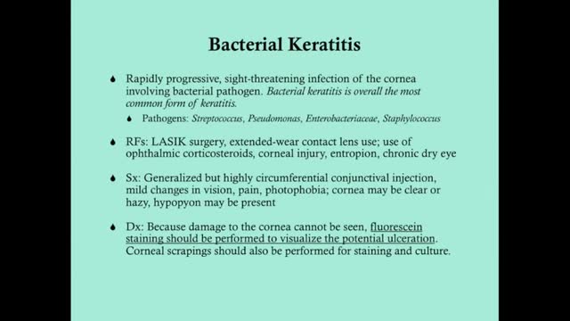
Keratitis is an inflammation of the cornea — the clear, dome-shaped tissue on the front of your eye that covers the pupil and iris. Keratitis is sometimes caused by an infection involving bacteria, viruses, fungi or parasites. Noninfectious keratitis can be caused by a minor injury, wearing your contact lenses too long or other noninfectious diseases. If you have eye redness or other symptoms of keratitis, make an appointment to see your doctor. With prompt attention, mild to moderate cases of keratitis can usually be effectively treated without loss of vision. If left untreated, or if an infection is severe, keratitis can lead to serious complications that may permanently damage your vision.
