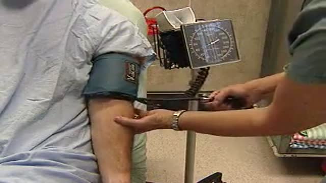
- Physical Examination
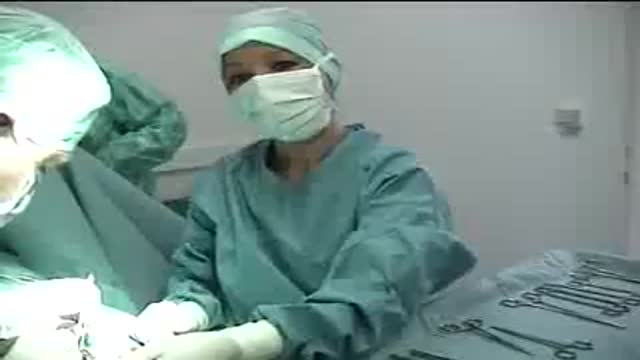
- Surgical Examination
- Ophthalmology
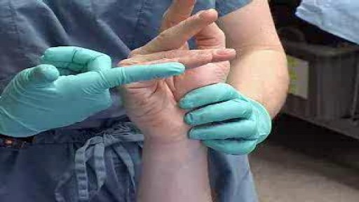
- Clinical Skills
- Orthopedics
- Surgery Videos
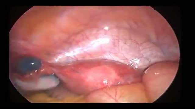
- Laparoscopy
- Pediatrics
- Funny Videos
- Cardiothoracic Surgery
- Nursing Videos
- Plastic Surgery
- Otorhinolaryngology
- Histology and Histopathology
- Neurosurgery
- Dermatology
- Pediatric Surgery
- Urology
- Dentistry
- Oncology and Cancers
- Anatomy Videos
- Health and Fitness
- Radiology
- Anaesthesia
- Physical Therapy
- Pharmacology
- Interventional Radiology
- Cardiology
- Endocrinology
- Gynecology
- Emergency Medicine
- Psychiatry and Psychology
- Childbirth Videos
- General Medical Videos
- Nephrology
- Physiology
- Diet and Food Health
- Diabetes Mellitus
- Neurology
- Women Health
- Osteoporosis
- Gastroenterology
- Pulmonology
- Hematology
- Rheumatology
- Toxicology
- Nuclear Medicine
- Infectious Diseases
- Vascular Disease
- Reproductive Health
- Burns and Wound Healing
- Other
Top videos
Inserteing a foley catheter in the male's urethra
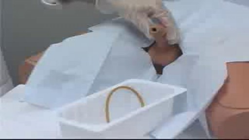
An old video showing how to give an enema
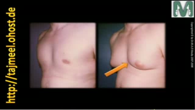
Gynecomastia means enlargement of male breast to resample female breast that is a common problem between males and causes many psychological problem
Dr. Mohamed El-Rouby
Consltant of Plastic surgery - Faculty of Medicine - Ain Shams University

Flexor compartment synovectomy in a patient with rheumatoid arthritis presenting with loss of finger movement and local pain due to synovitis. Performed at the Queen Victoria Hospital, East Grinstead.
Watch as Dr. Benjamin Carson performs risky brain surgery on young Payton to remove a brain tumor. Dr. Carson, director of pediatric neurosurgery, is just one of the many reasons why Johns Hopkins Children's Center was recently ranked #1 in neurology and neurosurgery in America's Best Children's Hospitals 2008
Adult circumcision video
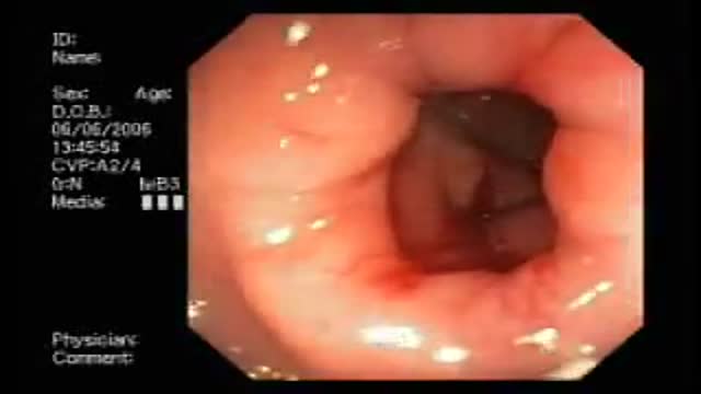
Bleeding from Duodenal Ulcer
Circumcision Video 3D
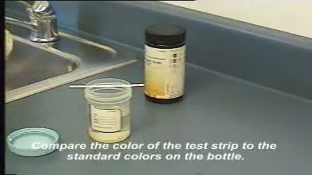
This video demonstrates how use a commercially-prepared "dip-stick" to test a random urine specimen for the presence of protein or glucose.
Fallopian Tube Diverticulus seen on Infertility workup Methylene Blue injected for tubal patency shows This. Edited by Dr Hemant Damle Prof & HOD Of Obs at SKN Medical College Pune India

This minimally invasive procedure for the replacement of a defective aortic valve has many advantages over traditional open heart surgery. ~ Detroit Medical Center

A video show phlebotomy tips

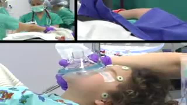
Patient Greg Grindley communicates with host Bryant Gumbel and his wife for the first time while undergoing deep brain stimulation surgery at University Hospital's Case Medical Center in Cleveland, Ohio.
➡ Subscribe: http://bit.ly/NatGeoSubscribe
About National Geographic:
National Geographic is the world's premium destination for science, exploration, and adventure. Through their world-class scientists, photographers, journalists, and filmmakers, Nat Geo gets you closer to the stories that matter and past the edge of what's possible.
Get More National Geographic:
Official Site: http://bit.ly/NatGeoOfficialSite
Facebook: http://bit.ly/FBNatGeo
Twitter: http://bit.ly/NatGeoTwitter
Instagram: http://bit.ly/NatGeoInsta
Greg's First In-Surgery Conversation | Brain Surgery Live
https://youtu.be/zvqV_2zncNU
National Geographic
https://www.youtube.com/natgeo
Some tips on obtaining venous blood samples.
Ulnar Gutter Cast
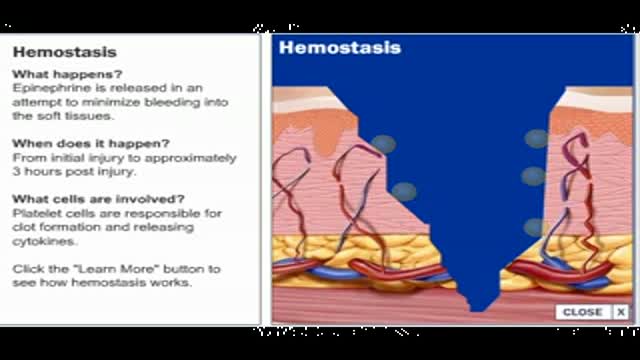
Wound healing, or wound repair, is the body's natural process of regenerating dermal and epidermal tissue. When an individual is wounded, a set of complex biochemical events takes place in a closely orchestrated cascade to repair the damage. These events overlap in time and may be artificially categorized into separate steps: the inflammatory, proliferative, and remodeling phases (Some authors consider healing to take place in four or more stages, by splitting different parts of inflammation or proliferation into separate steps.). In the inflammatory phase, bacteria and debris are phagocytized and removed, and factors are released that cause the migration and division of cells involved in the proliferative phase.

Una revision unica de fertilizacion, desarrollo embrionario y de los procedimientos llevados acabo durante un ciclo de fertilizacion invitro. Tome un tour virtual exclusivo de unos de los laboratorios de fertilizacion invitro mas avanzados del mundo y con tecnologia de punta en reproduccion asistida para que conozca con mas detalle como RMA de NY realiza estos procedimientos bajo control estricto de calidad.
Este video proporciona documentacion acerca de la aspiracion de ovulos, inseminacion de ovulos, desarrollo embrionario desde etapa de clivaje (2-3 dias) hasta etapa de blastocisto (5-6 dias), inyeccion intracitplasmatica de esperma (ICSI)), eclosion asistida, transferencia embrionaria y congelacion de embriones.
Mexico City
Dr. Benjamin Sandler
Reproductive Medicine Associates International
http://www.rmany.com/mexicointernatio...
Prolongacion Paseo de la Reforma 1232, Oficina 1213
Colonia Lomas de Bezares
Delegacion Miguel Hidalgo
Mexico, Distrito Federal 11910
Tel: 011-52-55-2167-2515
Fax: 011-52-55-2167-6434

A very funny video by Michael Moore showing a brief history of America
Medical Examination of the cranial nerves
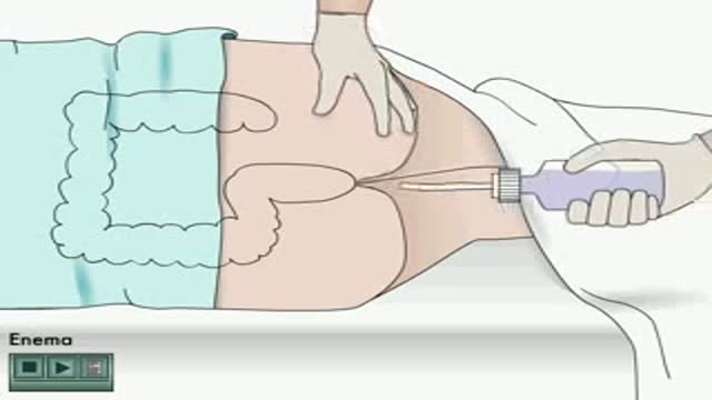
Enema how to apply Animation
