- Physical Examination
- Surgical Examination
- Ophthalmology
- Clinical Skills
- Orthopedics
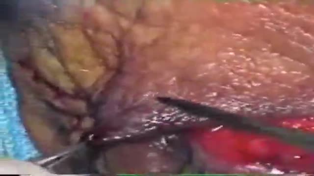
- Surgery Videos
- Laparoscopy
- Pediatrics
- Funny Videos
- Cardiothoracic Surgery
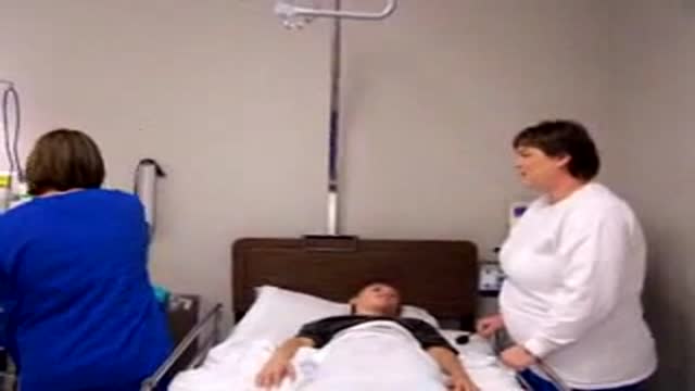
- Nursing Videos
- Plastic Surgery
- Otorhinolaryngology
- Histology and Histopathology
- Neurosurgery
- Dermatology
- Pediatric Surgery
- Urology
- Dentistry
- Oncology and Cancers
- Anatomy Videos
- Health and Fitness
- Radiology
- Anaesthesia
- Physical Therapy
- Pharmacology
- Interventional Radiology
- Cardiology
- Endocrinology
- Gynecology
- Emergency Medicine
- Psychiatry and Psychology
- Childbirth Videos
- General Medical Videos
- Nephrology
- Physiology
- Diet and Food Health
- Diabetes Mellitus
- Neurology
- Women Health
- Osteoporosis
- Gastroenterology
- Pulmonology
- Hematology
- Rheumatology
- Toxicology
- Nuclear Medicine
- Infectious Diseases
- Vascular Disease
- Reproductive Health
- Burns and Wound Healing
- Other
Top videos
Preventing Perineal Tears HD
Pediatric Febrile Seizures
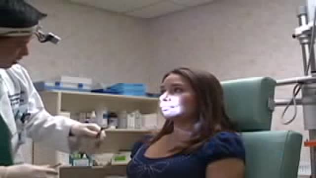
Stop Nose Bleeds by Cautery
Eye Lid Jones Procedure
Cleft Lip Surgery Millard Unilateral
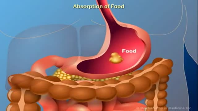
The Role of Insulin in the Human Body
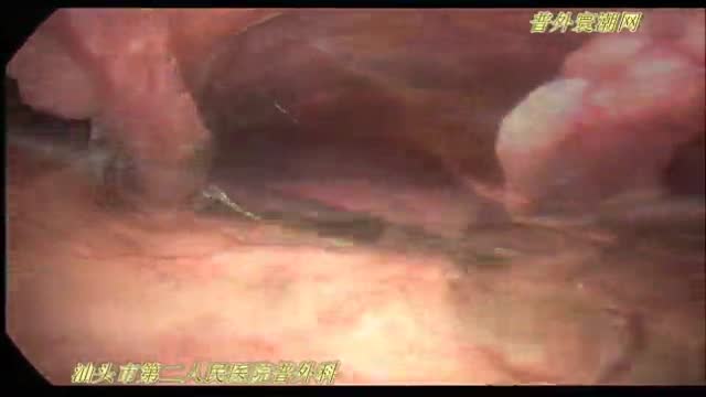
腹腔镜十二指肠球部溃疡穿孔修补术
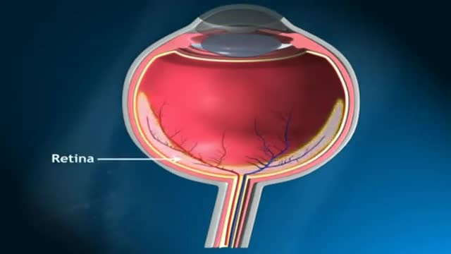
Glaucoma is called the silent thief of sight. It does not have symptoms during the early stages of the diseases and can make a patient blind over several years
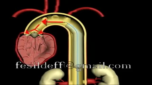
Intra Aortic Balloon Pump
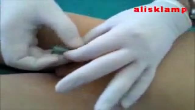
this video shows how the child circumcision is easy and safe with alisklamp

35 year old women with breathing difficulties for 6 months and feels like fluid is leaking down her front and back. Pain in thorax, lower back and pelvic. Weight loss. Was exposed to mold for a 2 years. Has a dog witch has persistent worm infection. Also been traveling out of the country.
High Blood Pressure Body Effects
Development and Maintenance of Bone
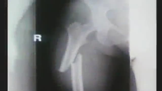
Treating osteoporosis with bisphosphonates, particularly for more than five years, has been linked to some side effects, including atypical femur fractures. Osteoporosis medications are supposed to prevent bone breaks. But if they are taken for too long, the opposite can happen. This video highlights what you need to know as a healthcare professional to educate patients
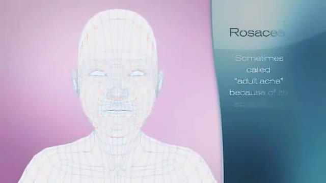
http://www.drmarylupo.com/ Rosacea is sometimes called 'adult acne', but it's not. Over 14million Americans have Rosacea. For most, Rosacea is an embarrassing cyclical condition, coming and going.
bilateral tubal ligation as modified Pomeroy technique during a C-Section
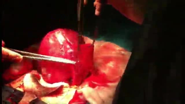
Uterine fibroids are the most common benign tumors and can affect one in three in Canada. While most fibroids are asymptomatic, they can cause heavy and painful periods, urinary frequency and urgency and pelvic discomfort and pain. A new treatment is available that doesn’t involve invasive surgery. With Fibristal, you can treat fibroids, relieve symptoms and finally live your life the way you want to!
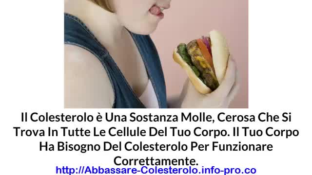
Come Alzare Il Colesterolo Buono, Colesterolo Hdl, Abbassare Il Colesterolo
http://abbassare-colesterolo.info-pro.co
COME EFFICACEMENTE ABBASSARE IL COLESTEROLO
senza prendere farmaci!
Il colesterolo è una sostanza molle, cerosa che si trova in tutte le cellule del tuo corpo. Il tuo corpo ha bisogno del colesterolo per funzionare correttamente. Il tuo corpo utilizza il colesterolo per tenere insieme le cellule. Inoltre il tuo corpo usa il colesterolo per creare gli ormoni, la vitamina D, e sostanze che aiutano a digerire gli alimenti.
Tuttavia, se troppo colesterolo entra nel sangue può causare problemi. Questo è noto come il colesterolo alto.
Se hai il colesterolo alto, e non fai nulla per abbassarlo, sarai ad un maggior rischio di gravi problemi di salute, come ad esempio un attacco di cuore o ictus. Pertanto, l'abbassamento del colesterolo è una questione importante per la salute generale di tutti.
Per saperne di più su come si può seguire un piano scientificamente provato per sconfiggere il colesterolo, visita il sito:
http://abbassare-colesterolo.info-pro.co
Clicca sul link sottostante per fare il check out
http://abbassare-colesterolo.info-pro.co
Iscriviti al nostro canale
http://www.youtube.com/user/viveresano01
https://www.youtube.com/watch?v=BWojp9nfdsU
Come Alzare Il Colesterolo Buono, Colesterolo Hdl, Abbassare Il Colesterolo,
colesterolo,
alimenti colesterolo,
colesterolo ldl basso,
colesterolo alimenti da evitare,
colesterolo e trigliceridi alti,
alimenti contro il colesterolo,
dieta x colesterolo,
cause colesterolo alto,
cosa mangiare per abbassare il colesterolo,
alimenti ricchi di colesterolo,
cibi contro il colesterolo

aasd
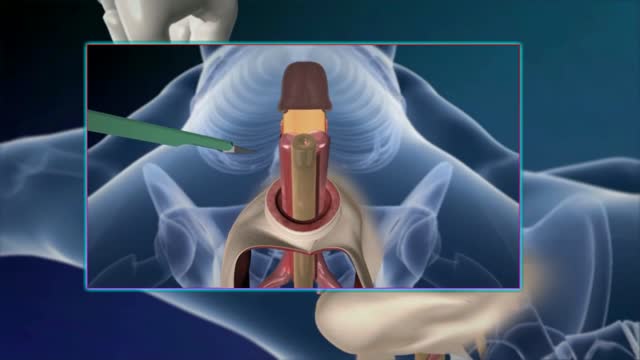
Sex reassignment surgery for male-to-female involves reshaping the male genitals into a form with the appearance of, and, as far as possible, the function of female genitalia. Prior to any surgeries, patients usually undergo hormone replacement therapy (HRT), and, depending on the age at which HRT begins, facial hair removal. There are associated surgeries patients may elect to, including facial feminization surgery, breast augmentation, and various other procedures
