- Physical Examination
- Surgical Examination
- Ophthalmology
- Clinical Skills
- Orthopedics
- Surgery Videos
- Laparoscopy
- Pediatrics
- Funny Videos
- Cardiothoracic Surgery
- Nursing Videos
- Plastic Surgery
- Otorhinolaryngology
- Histology and Histopathology
- Neurosurgery
- Dermatology
- Pediatric Surgery
- Urology
- Dentistry
- Oncology and Cancers
- Anatomy Videos
- Health and Fitness
- Radiology
- Anaesthesia
- Physical Therapy
- Pharmacology
- Interventional Radiology
- Cardiology
- Endocrinology
- Gynecology
- Emergency Medicine
- Psychiatry and Psychology
- Childbirth Videos
- General Medical Videos
- Nephrology
- Physiology
- Diet and Food Health
- Diabetes Mellitus
- Neurology
- Women Health
- Osteoporosis
- Gastroenterology
- Pulmonology
- Hematology
- Rheumatology
- Toxicology
- Nuclear Medicine
- Infectious Diseases
- Vascular Disease
- Reproductive Health
- Burns and Wound Healing
- Other
Top videos
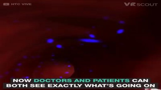
VR medical training takes you inside the human body.
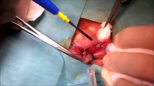
Watch that Hemorrhoid Medical Removal Surgery

Sialadenitis is an infection of the salivary glands. It is usually caused by a virus or bacteria . The parotid (in front of the ear) and submandibular (under the chin) glands are most commonly affected. Sialadenitis may be associated with pain, tenderness, redness, and gradual, localized swelling of the affected area.

Vaginal Yeast Infection
Watch that video of The World's Worst Spider Bites

A Hundred Orgasms A Day follow the story of 3 women who were tormented every hour of everyday with the need to have orgasm. This documentary explain how Persistent Sexual Arousal Syndrome or PSAS causes this unusual condition. PSAS is a little know neurological disorder where women have symptoms of continuous uncontrollable genital arousal. This condition is unrelated to any kind of sensations of sexual desire. PSAS was initially documented by Doctor Sandra Leiblum in mid 2001, just recently recognized as a unique syndrome in medical science which has a comparable equivalent progressively more claimed by men. A few physicians makes use of the name Persistent Sexual Arousal Syndrome to reference the disorder in women; some others look at the syndrome of priapism in adult males to be a similar disorder. Most importantly, it is really not connected with hyper-sexuality, also known as nymphomania. Both hyper-sexuality, and nymphomania are not known diagnosable health conditions. Not only is it very rare, the disorder is also seldom reported by affected individual who may think it is embarrassing.

Musculoskeletal Physical Examination Lecture

Gowers' sign is a medical sign that indicates weakness of the proximal muscles, namely those of the lower limb. The sign describes a patient that has to use his hands and arms to "walk" up his own body from a squatting position due to lack of hip and thigh muscle strength. It is named for William Richard Gowers. Gowers' sign is classically seen in Duchenne muscular dystrophy, but also presents itself in centronuclear myopathy, myotonic dystrophy and various other conditions associated with proximal muscle weakness. For this maneuver, the patient is placed on the floor away from any objects that could otherwise be used to pull oneself to a standing position. It is also used in testing paraplegia.

Doctors try to find a way for their patient suffering from a rare skin condition that causes her skin to blister and bleed if it touches anything, to attend her senior prom.
From Pure Genius Season 1 Episode 11 'Touch and Go' - James and Zoe try radical treatments on a girl with a rare skin disorder in an attempt to heal her in time for her prom; James considers an experimental cure for Louis Keating's GSS condition; Malik is jealous of James and Zoe's special connection.
Pure Genius (2016) A young tech-titan from Silicon Valley decides to build a hospital with a new-school approach to medicine and enlists a veteran surgeon who has a controversial past.
Watch full episodes of Pure Genius here: https://www.justwatch.com/uk/tv-series/pure-genius
Welcome to MD TV! A channel dedicated to your favourite medical dramas! Featuring iconic moments from House M.D., Chicago Med and more. Follow the professional and personal lives of the hospital staff, as you go a journey right from the very first doctor's call to the E.R and beyond. MD TV is packed full of drama, intrigue, and plenty of medical emergencies!
#MDTV #medicaldrama #medicaltvshow
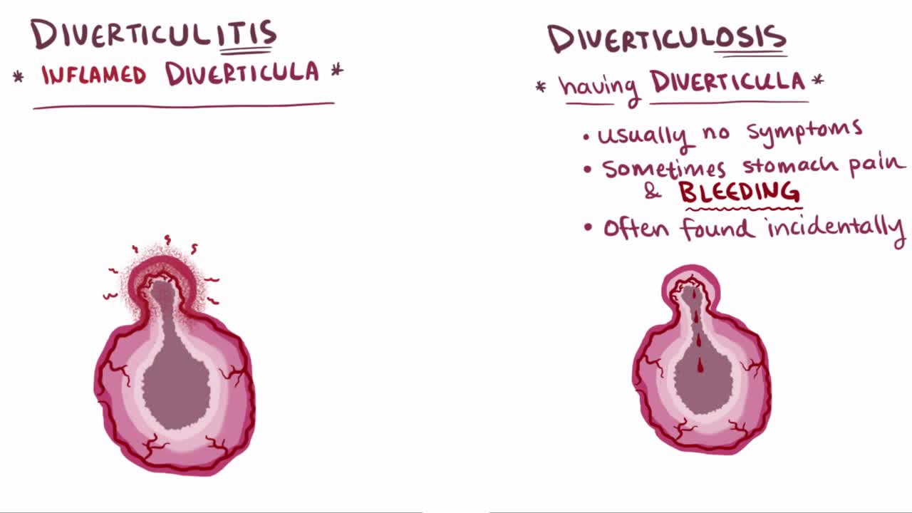
What are diverticula? Diverticula are outpouchings that most commonly happen in the sigmoid colon of the large intestine. The presence of a diverticulum is defined as diverticulosis, whereas diverticulitis describes an inflamed diverticulum
There’s a strange, mysterious world inside us, an alien-looking environment that turns the food we eat into nutrients that keep us alive. Michael Mosley swallows a camera to take a closer look.

► Sign up here and try our FREE content: http://lectur.io/freecontentyt
► If you’re an medical educator or faculty member, visit: http://lectur.io/medytb2u
This video “Connective Tissue” is part of the Lecturio course “Histology” ► WATCH the complete course on http://lectur.io/connectivetissue
► LEARN ABOUT:
- Cells and Basic Tissue
- Nerve Tissues
- Muscle Tissues
- Epithelial Tissues
- Connective Tissues
► THE PROF: Your lecturer is Professor Geoff Meyer. He is currently teaching at the School of Anatomy, Physiology and Human Biology at the University of Western Australia (UWA). As a leading anatomy and histology expert he is also coordinating the Federative International Program for Anatomical Terminologies (FIPAT) of the International Federation of Associations of Anatomists (IFAA). Besides medical research on the ovarian function, steroidogenesis, corpus luteum, angiogenesis, and microcirculation, Geoff Meyer’s research activities also focus on developing innovative, computer-aided learning and teaching tools. For his inventiveness, Geoff Meyer has received a number of awards, including the Australian University Teaching Award.
► LECTURIO is your single-point resource for medical school:
Study for your classes, USMLE Step 1, USMLE Step 2, MCAT or MBBS with video lectures by world-class professors, recall & USMLE-style questions and textbook articles. Create your free account now: http://lectur.io/connectivetissue
► INSTALL our free Lecturio app
iTunes Store: https://app.adjust.com/z21zrf
Play Store: https://app.adjust.com/b01fak
► READ TEXTBOOK ARTICLES related to this video:
Types of Tissue: Connective Tissue, Muscle Tissue, Epithelial Tissue, and Nervous Tissue
http://lectur.io/connectivetissuearticle
► SUBSCRIBE to our YouTube channel: http://lectur.io/subscribe
► WATCH MORE ON YOUTUBE: http://lectur.io/playlists
► LET’S CONNECT:
• Facebook: https://www.facebook.com/lectu....rio.medical.educatio
• Instagram: https://www.instagram.com/lecturio_medical_videos
• Pinterest: https://www.pinterest.de/lecturiomedical
• LinkedIn: https://www.linkedin.com/company/lecturio-medical/
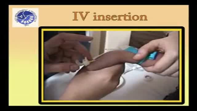
Pediatric IV insertion
Ganglion Cyst Removal
High volume sinus irrigation!
In today's video our patient is on the second stage of her breast reconstruction journey. Previously she had a mastectomy on the left side then we inserted a tissue expander to help stretch the breast tissue to create a pocket for the permanent breast implant that we are placing in today's video. On top of the breast implant we are grafting this patient's own fat into the breast to add a little extra volume and help it be more symmetrical with the other breast.
A video showing the technique of BoTox injection which is widely used by plastic surgeons to make wrinkles disappear
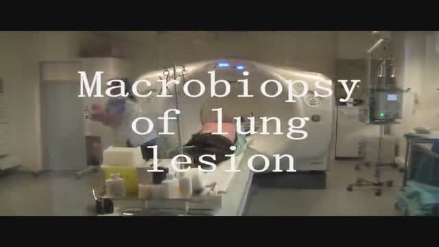
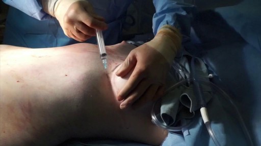
Spirotome macrobiopsy of a lung as a minimal invasive way to complete the diagnosis of lung lesions.

An antisperm antibody test looks for special proteins (antibodies) that fight against a man's sperm in blood, vaginal fluids, or semen. The test uses a sample of sperm and adds a substance that binds only to affected sperm. Semen can cause an immune system response in either the man's or woman's body. The antibodies can damage or kill sperm. If a high number of sperm antibodies come into contact with a man's sperm, it may be hard for the sperm to fertilize an egg. The couple has a hard time becoming pregnant. This is called immunologic infertility.
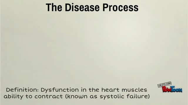
Dilated cardiomyopathy (DCM) is a condition in which the heart's ability to pump blood is decreased because the heart's main pumping chamber, the left ventricle, is enlarged and weakened.
