Top videos
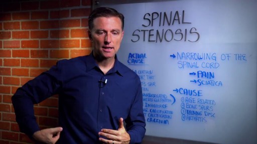
Spinal stenosis can put pressure on the spinal cord and the nerves within the spine. It commonly occurs in the neck and lower back. The condition is often caused by age-related wear and tear. Symptoms, if they occur, include pain, numbness, muscle weakness, and impaired bladder or bowel control. Treatment includes medication, physical therapy, and possibly surgery
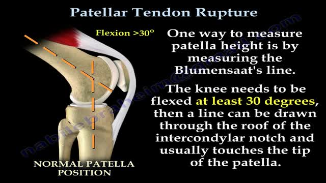
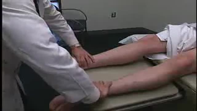
Patellar tendon rupture is a rupture of the tendon that connects the patella to the tibia. The superior portion of the patellar tendon attaches on the posterior portion of the patella, and the posterior portion of the patella tendon attaches to the tibial tubercle on the front of the tibia.
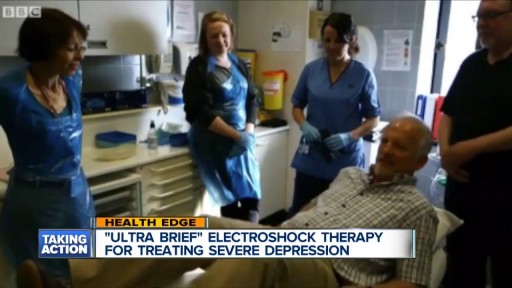
With ECT, electrodes are placed on the patient's scalp and a finely controlled electric current is applied while the patient is under general anesthesia. The current causes a brief seizure in the brain. ECT is one of the fastest ways to relieve symptoms in severely depressed or suicidal patients.
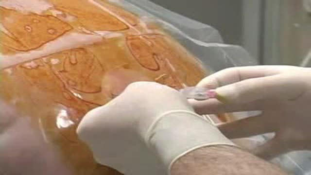
As you can see I access the left implant from the periareolar incisions which I made at the lower portion of the areola. As I entered the capsule and begin to remove the implant I noticed a lot of fluid surrounding the implant. Right away I know this is a rupture and that the mammogram was incorrect. Mammograms are very helpful in detecting cancer but often not ruptures. When implants rupture, it is important to have them replaced as soon as possible to avoid excessive scarring in the breasts. If too much scar tissue has accumulated around the deflated implant, it becomes difficult to create a normal breast shape in the future. Therefor know the signs of a ruptured implant such as, painful to touch, visible asymmetry or loss of integrity to the bag. For more information please visit: www.drlinder.com

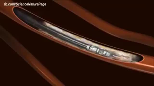
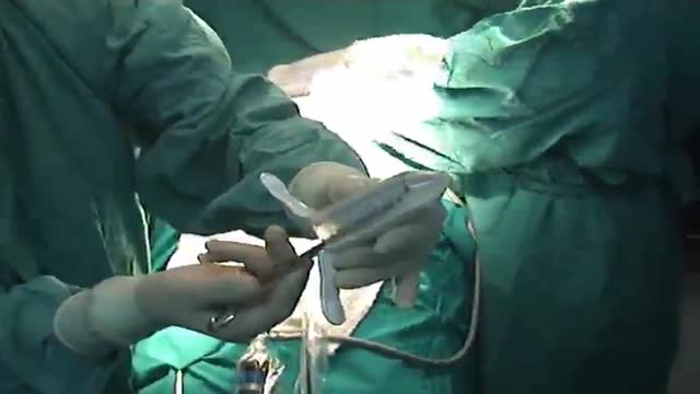
DMC Pediatric Heart Specialist Doctor Peter Karpawich is the first in the state to use minimally invasive surgery to repair a damaged pacemaker on a pediatric patient, helping her lead a more active, fulfilling lifestyle. ~ Detroit Medical Center
Cholesterol is a fat-like, waxy substance that can be found in all parts of your body. It helps your body make cell membranes, many hormones, and vitamin D. The cholesterol in your blood comes from two sources: the foods you eat and your liver. But your liver makes all the cholesterol your body needs.
Educational video of male patient receiving an anoscopy.

A video from Physical Exam Series of Loyola University Health System, Chicago showing the medical examination of the abdomen
Full examination of the female from head to toe by Loyola Medical School, Chicago. Part 3
Hemodialysis
Loyola Full Male Exam Part 4 A video from Loyola medical school, Chicago showing the full examination of the male
Paramedian Thoracic Epidural Anaesthesia
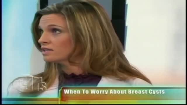
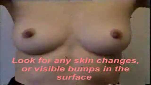
The lumps may be hard or rubbery and can appear as a single breast lump that may be large or small. Fibrocystic changes also can appear as thickening of the breast tissue. Fibrocystic changes can occur in one or both breasts and are the most common cause of benign breast lumps in women age 35 to 50.
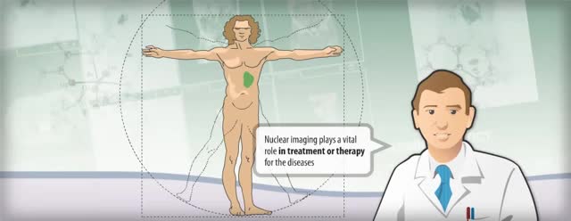
Nuclear medicine is a branch of medical imaging that uses small amounts of radioactive material to diagnose and determine the severity of or treat a variety of diseases, including many types of cancers, heart disease, gastrointestinal, endocrine, neurological disorders and other abnormalities within the body.
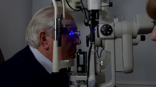
Eye Pressure Test
Endoscopic Atraumatic Coronary Artery Bypass EndoACA
Brought to you by http://nursing-resource.com
Internal hemorrhoids and loose rectal mucosa may block the exposure during the purse string suturing in stapled hemorrhooidopexy, and this may cause some complications. To retract the prolapsing rectal mucosa we modified the purse string anoscope of the PPH01 kit (Ethicon-Endosurgery, Cincinnati, O...H, USA) and produced a special anoscope. The open part of the purse string suture anoscope is covered by transparent acrylic (Orthoacryl�, Dentaurum, Pforzheim, Germany). The covering material had complete cylindrical outer and inner surfaces and was thin enough to let the anoscope easily rotate in the anal dilator and to let the 26 mm curved, round bodied needle of the 2/0 polypropilene suture move in the anoscope. A window, 3 cm long and 3-4 mm wide, was opened at the angled part of the anoscope 2 cm to the tip of the anoscope. This special anoscope was used for the purse string suture during stapled hemorrhoidopexy procedure in five patients. No postoperative complications, early or late, were encountered, and we propose that stapled hemorrhoidopexy procedure can be applied more easily by using this special anoscope.

If you have an active lifestyle or are often on the go with work, travel or family, then peritoneal dialysis at home may be the right choice. Home peritoneal dialysis offers additional freedom and flexibility as a treatment option that’s closest to natural kidney function and may require fewer dietary restrictions and medications. To learn more about Home PD, visit https://www.FreseniusKidneyCar....e.com/ckd-treatment/
Subscribe to Our YouTube Channel
Subscribe Here: https://www.youtube.com/c/Fres....eniusKidneyCareOffic
Find Fresenius Kidney Care Online at:
Website: https://www.freseniuskidneycare.com/
Facebook:@FreseniusKidneyCare
https://www.facebook.com/FreseniusKidneyCare/
Twitter: @FreseniusKC
https://twitter.com/freseniuskc
Pinterest: Fresenius Kidney Care
https://www.pinterest.com/FreseniusKidneyCarePins/
