- Physical Examination
- Surgical Examination
- Ophthalmology
- Clinical Skills
- Orthopedics
- Surgery Videos
- Laparoscopy
- Pediatrics
- Funny Videos
- Cardiothoracic Surgery
- Nursing Videos
- Plastic Surgery
- Otorhinolaryngology
- Histology and Histopathology
- Neurosurgery
- Dermatology
- Pediatric Surgery
- Urology
- Dentistry
- Oncology and Cancers
- Anatomy Videos
- Health and Fitness
- Radiology
- Anaesthesia
- Physical Therapy
- Pharmacology
- Interventional Radiology
- Cardiology
- Endocrinology
- Gynecology
- Emergency Medicine
- Psychiatry and Psychology
- Childbirth Videos
- General Medical Videos
- Nephrology
- Physiology
- Diet and Food Health
- Diabetes Mellitus
- Neurology
- Women Health
- Osteoporosis
- Gastroenterology
- Pulmonology
- Hematology
- Rheumatology
- Toxicology
- Nuclear Medicine
- Infectious Diseases
- Vascular Disease
- Reproductive Health
- Burns and Wound Healing
- Other
Top videos

Nosebleeds common. Most often they are a nuisance and not a true medical problem. But they can be both. Nosebleed care Sit upright and lean forward. By remaining upright, you reduce blood pressure in the veins of your nose. This discourages further bleeding. Sitting forward will help you avoid swallowing blood, which can irritate your stomach. Pinch your nose. Use your thumb and index finger to pinch your nostrils shut. Breathe through your mouth. Continue to pinch for five to 10 minutes. Pinching sends pressure to the bleeding point on the nasal septum and often stops the flow of blood. To prevent re-bleeding, don't pick or blow your nose and don't bend down for several hours after the bleeding episode. During this time remember to keep your head higher than the level of your heart. If re-bleeding occurs, blow out forcefully to clear your nose of blood clots and spray both sides of your nose with a decongestant nasal spray containing oxymetazoline (Afrin, Mucinex Moisture Smart, others). Pinch your nose again as described above and call your doctor. When to seek emergency care The bleeding lasts for more than 20 minutes The nosebleed follows an accident, a fall or an injury to your head, including a punch in the face that may have broken your nose
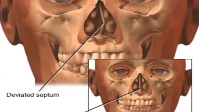
Nosebleeds are common. Most often they are a nuisance and not a true medical problem. But they can be both. Nosebleed care Sit upright and lean forward. By remaining upright, you reduce blood pressure in the veins of your nose. This discourages further bleeding. Sitting forward will help you avoid swallowing blood, which can irritate your stomach. Pinch your nose. Use your thumb and index finger to pinch your nostrils shut. Breathe through your mouth. Continue to pinch for five to 10 minutes. Pinching sends pressure to the bleeding point on the nasal septum and often stops the flow of blood. To prevent re-bleeding, don't pick or blow your nose and don't bend down for several hours after the bleeding episode. During this time remember to keep your head higher than the level of your heart. If re-bleeding occurs, blow out forcefully to clear your nose of blood clots and spray both sides of your nose with a decongestant nasal spray containing oxymetazoline (Afrin, Mucinex Moisture Smart, others). Pinch your nose again as described above and call your doctor. When to seek emergency care The bleeding lasts for more than 20 minutes The nosebleed follows an accident, a fall or an injury to your head, including a punch in the face that may have broken your nose
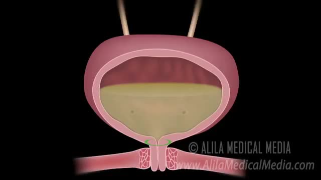
Urinary incontinence is the unintentional loss of urine. Stress incontinence happens when physical movement or activity — such as coughing, sneezing, running or heavy lifting — puts pressure (stress) on your bladder. Stress incontinence is not related to psychological stress. Stress incontinence differs from urge incontinence, which is the unintentional loss of urine caused by the bladder muscle contracting, usually associated with a sense of urgency. Stress incontinence is much more common in women than men. If you have stress incontinence, you may feel embarrassed, isolate yourself, or limit your work and social life, especially exercise and leisure activities. With treatment, you'll likely be able to manage stress incontinence and improve your overall well-being.
Urinary incontinence isn't a disease, it's a symptom. It can be caused by everyday habits, underlying medical conditions or physical problems. A thorough evaluation by your doctor can help determine what's behind your incontinence. Temporary urinary incontinence Certain drinks, foods and medications can act as diuretics — stimulating your bladder and increasing your volume of urine. They include: Alcohol Caffeine Decaffeinated tea and coffee Carbonated drinks Artificial sweeteners Corn syrup Foods that are high in spice, sugar or acid, especially citrus fruits Heart and blood pressure medications, sedatives, and muscle relaxants Large doses of vitamins B or C Urinary incontinence also may be caused by an easily treatable medical condition, such as: Urinary tract infection. Infections can irritate your bladder, causing you to have strong urges to urinate, and sometimes incontinence. Other signs and symptoms of urinary tract infection include a burning sensation when you urinate and foul-smelling urine. Constipation. The rectum is located near the bladder and shares many of the same nerves. Hard, compacted stool in your rectum causes these nerves to be overactive and increase urinary frequency. Persistent urinary incontinence Urinary incontinence can also be a persistent condition caused by underlying physical problems or changes, including: Pregnancy. Hormonal changes and the increased weight of the uterus can lead to stress incontinence. Childbirth. Vaginal delivery can weaken muscles needed for bladder control and also damage bladder nerves and supportive tissue, leading to a dropped (prolapsed) pelvic floor. With prolapse, the bladder, uterus, rectum or small intestine can get pushed down from the usual position and protrude into the vagina. Such protrusions can be associated with incontinence. Changes with age. Aging of the bladder muscle can decrease the bladder's capacity to store urine. Menopause. After menopause women produce less estrogen, a hormone that helps keep the lining of the bladder and urethra healthy. Deterioration of these tissues can aggravate incontinence. Hysterectomy. In women, the bladder and uterus are supported by many of the same muscles and ligaments. Any surgery that involves a woman's reproductive system, including removal of the uterus, may damage the supporting pelvic floor muscles, which can lead to incontinence. Enlarged prostate. Especially in older men, incontinence often stems from enlargement of the prostate gland, a condition known as benign prostatic hyperplasia. Prostate cancer. In men, stress incontinence or urge incontinence can be associated with untreated prostate cancer. But more often, incontinence is a side effect of treatments for prostate cancer. Obstruction. A tumor anywhere along your urinary tract can block the normal flow of urine, leading to overflow incontinence. Urinary stones — hard, stone-like masses that form in the bladder — sometimes cause urine leakage. Neurological disorders. Multiple sclerosis, Parkinson's disease, stroke, a brain tumor or a spinal injury can interfere with nerve signals involved in bladder control, causing urinary incontinence.

Best Position for Getting Pregnant Fast

Osteoporosis is a disease that weakens bones over time. Because of this, it puts you at risk for breaking a bone. Postmenopausal osteoporosis is the most common form of osteoporosis. It affects many women after menopause, leaving some women at high risk for fracture. - See more at: https://www.prolia.com/postmenopausal-osteoporosis/deeper-look/?WT.z_co=A&WT.z_in=PMO&WT.z_ch=PDS&WT.z_st=Site1&WT.z_mt=Broad&WT.z_pdskw=www%20osteoporosis&WT.z_ag=Osteoporosis%20Information%20-%20Broad&WT.z_se=G&WT.srch=1&WT.z_prm=DSE__&WT.mc_id=A_PMO_PDS_G_DSE___Broad_www%20osteoporosis_Osteoporosis%20Information%20-%20Broad#sthash.x5Ys3Now.dpuf

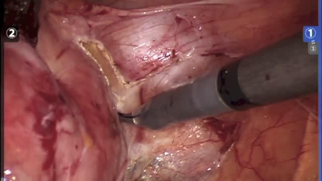
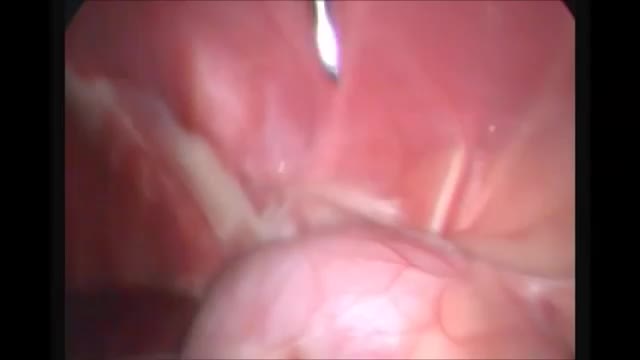
procedure is usually done in the hospital or outpatient surgical center under general anesthesia (while you are asleep and pain-free). The procedure is performed in the following way: The surgeon makes a small cut (incision) below the belly button (navel). A needle or tube is inserted into the incision. Carbon dioxide gas is passed into the abdomen through the needle or tube. The gas helps expand the area, giving the surgeon more room to work, and helping the surgeon see the organs more clearly. A tube is placed through the cut in your abdomen. A tiny video camera (laparoscope) goes through this tube and is used to see the inside of your pelvis and abdomen. More small cuts may be made if other instruments are needed to get a better view of certain organs. If you are having gynecologic laparoscopy, dye may be injected into your cervix area so the surgeon can view your fallopian tubes. After the exam, the gas, laparoscope, and instruments are removed, and the cuts are closed. You will have bandages over those areas.
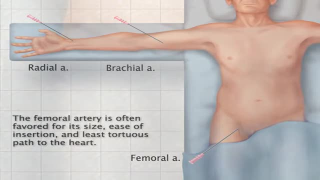
Transfemoral Cardiac Catheterization
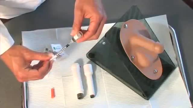
Penile Injection Therapy
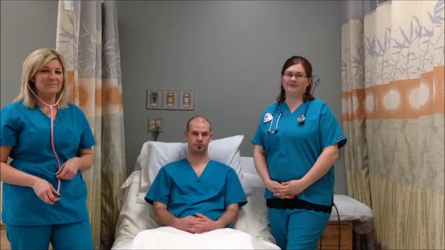
Breath sounds can be either normal or abnormal. These sounds come from the lungs when you breathe in or out. These sounds can be heard using a stethoscope or simply when breathing. Abnormal breath sounds can indicate a lung problem, such as: an obstruction inflammation an infection fluid in the lungs asthma Listening to breath sounds is an important part of diagnosing many different medical conditions.

Heart sounds are the noises generated by the beating heart and the resultant flow of blood through it. Specifically, the sounds reflect the turbulence created when the heart valves snap shut. In cardiac auscultation, an examiner may use a stethoscope to listen for these unique and distinct sounds that provide important auditory data regarding the condition of the heart. In healthy adults, there are two normal heart sounds often described as a lub and a dub (or dup), that occur in sequence with each heartbeat. These are the first heart sound (S1) and second heart sound (S2), produced by the closing of the atrioventricular valves and semilunar valves, respectively. In addition to these normal sounds, a variety of other sounds may be present including heart murmurs, adventitious sounds, and gallop rhythms S3 and S4.
Ganglion cysts are noncancerous lumps that most commonly develop along the tendons or joints of your wrists or hands. They also may occur in the ankles and feet. Ganglion cysts are typically round or oval and are filled with a jellylike fluid. Small ganglion cysts can be pea-sized, while larger ones can be around an inch (2.5 centimeters) in diameter. Ganglion cysts can be painful if they press on a nearby nerve. Their location can sometimes interfere with joint movement. If your ganglion cyst is causing you problems, your doctor may suggest trying to drain the cyst with a needle. Removing the cyst surgically also is an option. But if you have no symptoms, no treatment is necessary. In many cases, the cysts go away on their own.
HYSTERECTOMY RECOVERY: ALL PROCEDURES ARE NOT CREATED EQUAL Too often, women are only given the option of an open hysterectomy for conditions like large fibroids or an enlarged uterus. Surgical techniques have evolved in the last decade, but across the United States, the number of women still having open hysterectomy procedures is unnecessarily staggering. Robotic procedures are becoming more common as hospitals invest nearly $2 million in the machine. While the robot does allow surgeons who are not necessarily trained in laparoscopic procedures to perform a more minimally invasive surgery, tools cannot replace skill. There is no added benefit to the patient and the surgery can cost on average up to $2,000 more than other laparoscopic options, and in some cases much higher.
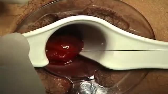
A surgeon begins the PPH stapled hemorrhoidectomy by inserting a circular anal dilator and obturator into the anal canal and then securing the dilator in place with four sutures. The surgeon then inserts a PPH anoscope into the obturator. Next, he places a circumferential purse-string suture of 2-0 Monocryl on a UR-6 needle 4 cm proximal to the dentate line. The surgeon opens a PPH stapler and places its anvil across the purse string. The stapler is then closed and fired; it is held closed for two minutes to improve hemostasis. Prior to firing the stapler in a female patient, the surgeon places a gloved finger in the vagina to ensure the vaginal mucosa and rectal-vaginal septum are not trapped within the jaws of the closed stapler. The surgeon then opens and removes the stapler.
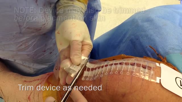
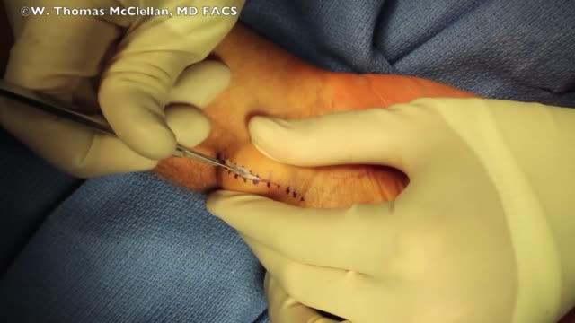
Zip Surgical Skin Closure

Red eyes usually are caused by allergy, eye fatigue, over-wearing contact lenses or common eye infections such as pink eye (conjunctivitis). However, redness of the eye sometimes can signal a more serious eye condition or disease, such as uveitis or glaucoma.

Reactive arthritis can affect the heels, toes, fingers, low back, and joints, especially of the knees or ankles. Though it often goes away on its own, reactive arthritis can be prolonged and severe enough to require seeing a specialist. Effective treatment is available for reactive arthritis. Reactive arthritis tends to occur most often in men between ages 20 and 50. Most cases of reactive arthritis appear as a short episode. Occasionally, it becomes chronic. Reactive arthritis is a painful form of inflammatory arthritis (joint disease due to inflammation). It occurs in reaction to an infection by certain bacteria. Most often, these bacteria are in the genitals (Chlamydia trachomatis) or the bowel (Campylobacter, Salmonella, Shigella and Yersinia). Chlamydia most often transmits by sex. It often has no symptoms, but can cause a pus-like or watery discharge from the genitals. The bowel bacteria can cause diarrhea. If you develop arthritis within one month of diarrhea or a genital infection – especially with a discharge – see a health care provider. You may have reactive arthritis. - See more at: http://www.rheumatology.org/I-Am-A/Patient-Caregiver/Diseases-Conditions/Reactive-Arthritis#sthash.VNgDSOOY.dpuf

Remove Acne Marks
A renal biopsy is a procedure used to extract kidney tissue for laboratory analysis. The word “renal” describes the kidneys. A renal biopsy is also called a kidney biopsy. The test helps your doctor identify the type of kidney disease you have, how severe it is, and the best treatment for it.

Sickle cell anemia causes pain, fatigue and delayed growth, all because of a lack of enough healthy red blood cells. And yet genetic mutations that cause it — recessive genes for the oxygen-carrying hemoglobin protein — have survived natural selection because they also seem to provide a natural defense against malaria.
