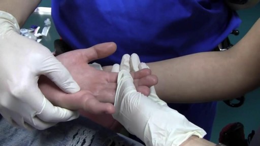
- Physical Examination
- Surgical Examination
- Ophthalmology
- Clinical Skills
- Orthopedics
- Surgery Videos
- Laparoscopy
- Pediatrics
- Funny Videos
- Cardiothoracic Surgery
- Nursing Videos
- Plastic Surgery
- Otorhinolaryngology
- Histology and Histopathology
- Neurosurgery
- Dermatology
- Pediatric Surgery
- Urology
- Dentistry
- Oncology and Cancers
- Anatomy Videos
- Health and Fitness
- Radiology
- Anaesthesia
- Physical Therapy
- Pharmacology
- Interventional Radiology
- Cardiology
- Endocrinology
- Gynecology
- Emergency Medicine
- Psychiatry and Psychology
- Childbirth Videos
- General Medical Videos
- Nephrology
- Physiology
- Diet and Food Health
- Diabetes Mellitus
- Neurology
- Women Health
- Osteoporosis
- Gastroenterology
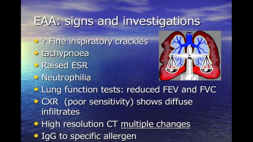
- Pulmonology
- Hematology
- Rheumatology
- Toxicology
- Nuclear Medicine
- Infectious Diseases
- Vascular Disease
- Reproductive Health
- Burns and Wound Healing
- Other
Top videos
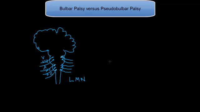
This tutorial explains the difference in mechanisms between the 2 palsies. Bulbar palsy is a lower motor neuron condition and pseudobulbar palsy is an upper motor neuron condidtion.
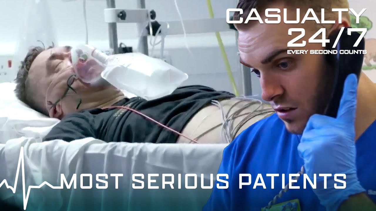
In this compilation, Barnsley Hospital is facing a very busy day with a high number of patients being treated, the doctors and nurses face some of their toughest shifts when they treat critical patients and rare illnesses as well as making tough decisions.
⌚️Timecodes:
00:00 Season 2 Episode 1
08:56 Season 4 Episode 1
16:53 Season 3 Episode 10
30:36 Season 3 Episode 13
37:45 Season 2 Episode 9
46:51 Season 1 Episode 2
52:52 Season 1 Episode 3
58:02 Season 2 Episode 2
01:09:39 Season 2 Episode 11
01:18:37 Season 2 episode 12
🟦 Click Link below to subscribe: https://www.youtube.com/channe....l/UCHPgATT2HtFrxmueq
About Casualty 24/7:
Casualty 24/7 shows how the doors of Barnsley A&E department are open every hour, of every day. They allow a peek inside their medical emergency teams, and how they deal with critical situations revolving around people's lives and illnesses. The team are close-knit and exchange typical Yorkshire humour to get them through their often long and tough days.
Watch our playlists:
🔵 Season 1 Full Episodes | Casualty 24/7:
https://www.youtube.com/playli....st?list=PLWrY8x74oDM
🔵 Season 2 Full Episodes | Casualty 24/7:
https://www.youtube.com/playli....st?list=PLWrY8x74oDM
🔵 Season 3 Full Episodes | Casualty 24/7:
https://www.youtube.com/playli....st?list=PLWrY8x74oDM
🔵 Season 4 Full Episodes | Casualty 24/7:
https://www.youtube.com/playli....st?list=PLWrY8x74oDM
🔵 Compilation Videos of Casualty 24/7:
https://www.youtube.com/playli....st?list=PLWrY8x74oDM
#SeriousIllness #Casualty247 #EmergencyServices #AandE #BHNFT #OurFutureSouthYorkshire
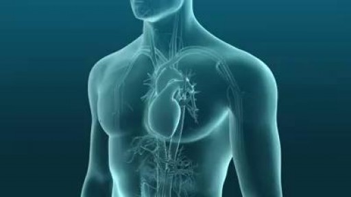
A ventricular septal defect (VSD) is an opening or hole in the wall that separates the two lower chambers of the heart. This wall is called the ventricular septum. The hole causes oxygen-rich blood to leak from the left side of the heart to the right side. This causes extra work for the right side of the heart, since more blood than necessary is flowing through the right ventricle to the lungs.
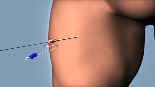
The fascinating way doctors insert a stent when there is plaque buildup in an artery.

Hemorrhoids (HEM-uh-roids), also called piles, are swollen veins in your anus and lower rectum, similar to varicose veins. Hemorrhoids have a number of causes, although often the cause is unknown. They may result from straining during bowel movements or from the increased pressure on these veins during pregnancy.
Occupational respiratory disease is any lung condition you get at work. Certain workplaces lend themselves to disease. The most common are coalmines and factories or areas with high amounts of toxins. These include asbestos and silica dust, as well as smoke, fumes, gases, and other particles. Types of occupational respiratory disease include: coal workers’ pneumoconiosis, also known as Black Lung Disease asbestosis silicosis farmers’ lung, also known as allergic alveolitis. It also includes forms of asthma, bronchitis, or emphysema.
Finger metacarpophalangeal (MCP) joint collateral ligament sprains should not be overtreated. First-degree sprains may require a brief period of protection, usually consisting of buddy taping for 2-3 weeks. Second-degree sprains are immobilized in mid flexion for 3 weeks. Finger MCP joint hyperextension injuries may be treated by gently flexing the proximal phalanx and immobilizing the MCP joint in 30° of flexion for 2-3 weeks. A dorsal extension-block splint protects the healing volar plate while allowing active flexion of the finger. Early protected motion minimizes postinjury stiffness. Thumb MCP joint hyperextension injuries ("locked MCP joint") are immobilized in 20° MCP joint flexion for 3 weeks.
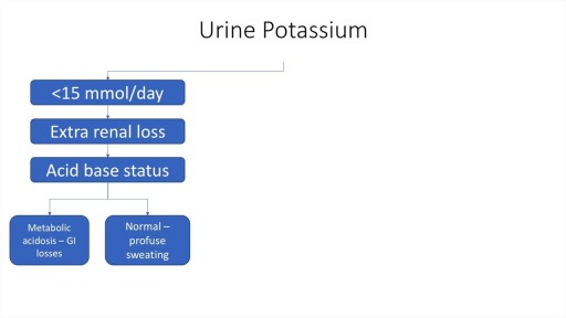
A step by step approach to Hypokalaemia including causes, diagnosis and management.

Dr. Carlos Benitez guides us through ultrasound images of the knee and how to identify knee injuries.
Watch that video to know How To Firm And Lift Your Sagging Chest Naturally
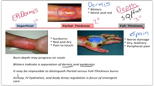
Burns are classified as first-, second-, or third-degree, depending on how deep and severe they penetrate the skin's surface. First-degree burns affect only the epidermis, or outer layer of skin. The burn site is red, painful, dry, and with no blisters. Mild sunburn is an example.
How Does a Bone Heal? All broken bones go through the same healing process. This is true whether a bone has been cut as part of a surgical procedure or fractured through an injury. The bone healing process has three overlapping stages: inflammation, bone production and bone remodeling. Inflammation starts immediately after the bone is fractured and lasts for several days. When the bone is fractured, there is bleeding into the area, leading to inflammation and clotting of blood at the fracture site. This provides the initial structural stability and framework for producing new bone. Diagram of inflammation in a fractured bone Bone production begins when the clotted blood formed by inflammation is replaced with fibrous tissue and cartilage (known as soft callus). As healing progresses, the soft callus is replaced with hard bone (known as hard callus), which is visible on x-rays several weeks after the fracture. Bone remodeling, the final phase of bone healing, goes on for several months. In remodeling, bone continues to form and becomes compact, returning to its original shape. In addition, blood circulation in the area improves. Once adequate bone healing has occurred, weightbearing (such as standing or walking) encourages bone remodeling.
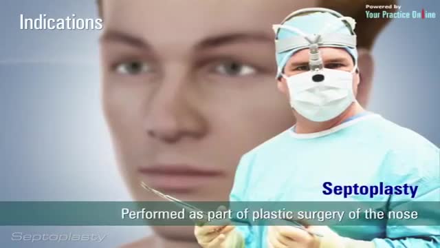
Septoplasty (SEP-toe-plas-tee) is a surgical procedure to correct a deviated septum — a displacement of the bone and cartilage that divides your two nostrils. During septoplasty, your nasal septum is straightened and repositioned in the middle of your nose.
Psychotic Depression Information

For more information:
http://www.7activestudio.com
info@7activestudio.com
http://www.7activemedical.com/
info@7activemedical.com
7activestudio@gmail.com
Contact: +91- 9700061777,
+91- 9100061777
7 Active Technology Solutions Pvt.Ltd. is an educational 3D digital content provider for K-12. We also customize the content as per your requirement for companies platform providers colleges etc . 7 Active driving force "The Joy of Happy Learning" -- is what makes difference from other digital content providers. We consider Student needs, Lecturer needs and College needs in designing the 3D & 2D Animated Video Lectures. We are carrying a huge 3D Digital Library ready to use.
Kidney is most essential organ to remove nitrogenous waste materials from the body. Kidney was damaged by several human activities leads to kidney failure. Once it is damaged it cannot perform basic functions. To overcome this problem one of the best method we follows called hemodialysis. Hemodialysis is a process of removing of nitrogenous waste materials and excess fluids from the blood (collecting from arteries) through tubes containing semi permeable linings in the dialyzer and sending purified blood to the patient's body through veins. It covers the process of hemodialysis in step wise manner. Hemodialysis only performs some basic functions not all those which are performed by natural kidney like reabsorption etc..
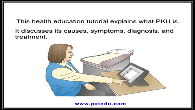
PKU is inherited in families in an autosomal recessive pattern. Autosomal recessive inheritance means that a person has two copies of the gene that is altered. Usually, each parent of an individual who has PKU carries one copy of the altered gene. ... Gene alterations (mutations) in the PAH gene cause PKU.
Head Eye and ENT Physical Examination
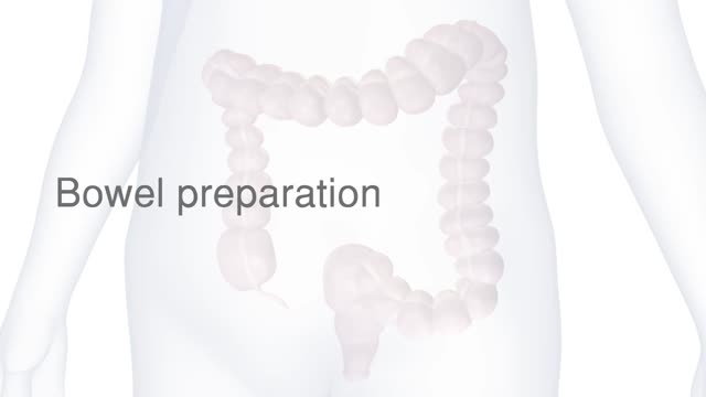
The camera sends images to an external monitor so the doctor can study the inside of your colon. The doctor can also insert instruments through the channel to take tissue samples (biopsies) or remove polyps or other areas of abnormal tissue. A colonoscopy typically takes about 20 minutes to an hour.
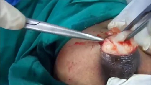
Different types of Abscess- Drainage and Aspiration of Pus.
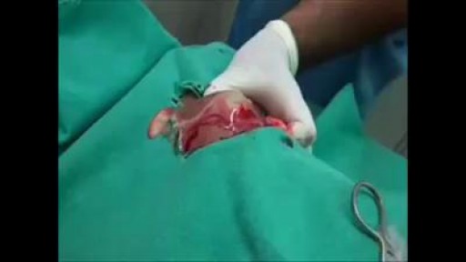
Watch that A Big Size Fibroadenoma Removal Under Local Anesthesia
