- Physical Examination
- Surgical Examination
- Ophthalmology
- Clinical Skills
- Orthopedics
- Surgery Videos
- Laparoscopy
- Pediatrics
- Funny Videos
- Cardiothoracic Surgery
- Nursing Videos
- Plastic Surgery
- Otorhinolaryngology
- Histology and Histopathology
- Neurosurgery
- Dermatology
- Pediatric Surgery
- Urology
- Dentistry
- Oncology and Cancers
- Anatomy Videos
- Health and Fitness
- Radiology
- Anaesthesia
- Physical Therapy
- Pharmacology
- Interventional Radiology
- Cardiology
- Endocrinology
- Gynecology
- Emergency Medicine
- Psychiatry and Psychology
- Childbirth Videos
- General Medical Videos
- Nephrology
- Physiology
- Diet and Food Health
- Diabetes Mellitus
- Neurology
- Women Health
- Osteoporosis
- Gastroenterology
- Pulmonology
- Hematology
- Rheumatology
- Toxicology
- Nuclear Medicine
- Infectious Diseases
- Vascular Disease
- Reproductive Health
- Burns and Wound Healing
- Other
Top videos
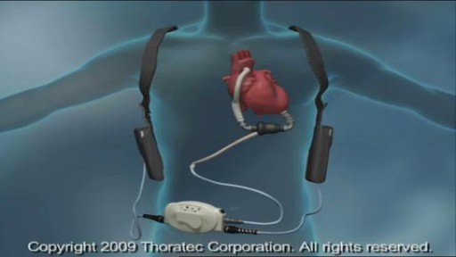
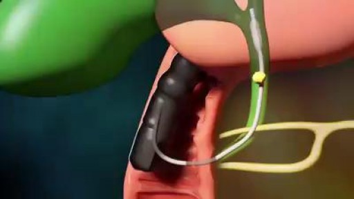
A ventricular assist device (VAD) — also known as a mechanical circulatory support device — is an implantable mechanical pump that helps pump blood from the lower chambers of your heart (the ventricles) to the rest of your body. A VAD is used in people who have weakened hearts or heart failure. Although a VAD can be placed in the left, right or both ventricles of your heart, it is most frequently used in the left ventricle. When placed in the left ventricle it is called a left ventricular assist device (LVAD). You may have a VAD implanted while you wait for a heart transplant or for your heart to become strong enough to effectively pump blood on its own. Your doctor may also recommend having a VAD implanted as a long-term treatment if you have heart failure and you're not a good candidate for a heart transplant.

Histology of Colon

Don't cleanse your contact lenses with tap water.
There’s a strange, mysterious world inside us, an alien-looking environment that turns the food we eat into nutrients that keep us alive. Michael Mosley swallows a camera to take a closer look.
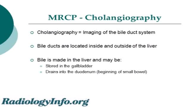
An MRCP scan is a scan that uses magnetic resonance imaging (MRI) to produce pictures of the liver, bile ducts, gallbladder and pancreas. Note: the information below is a general guide only. The arrangements,and the way tests are performed, may vary between different hospitals.
Spermicide is a birth control method that contains chemicals that stop sperm from moving. Spermicides are available in different forms, including creams, film, foams, gels, and suppositories. Spermicide can be used alone, or it can be used with other birth control methods to make them more effective. It is always used with the diaphragm and cervical cap.

Ways to Help Pregnant Women Dilate HD

Not sure what to expect with your child's upcoming surgery at Wesley Children's Hospital? This guided tour will walk you through the process to make both patients and families feel as comfortable as possible.

Colorectal cancer (also known as colon cancer, rectal cancer or bowel cancer) is the development of cancer in the colon or rectum (parts of the large intestine). It is due to the abnormal growth of cells that have the ability to invade or spread to other parts of the body. People with HNPCC tend to develop colon cancer before age 50. Familial adenomatous polyposis (FAP). FAP is a rare disorder that causes you to develop thousands of polyps in the lining of your colon and rectum. People with untreated FAP have a greatly increased risk of developing colon cancer before age 40.

Neck Examination - Cervical Spine Assessment - Clinical Skills - Dr Gill
Compose a new pain within athletes is cervical spine discomfort, thankfully in the vast majority of cases when the neck is examined the cause of the neck pain is found to be muscular.
However, pain can also refer from the neck to the arm, in which case it is important to be able to assess for cervical radiculopathy prior to gaining more information which may indicate an MRI is needed
We assess for radiculopathy by doing Spurling's test, an often overlooked part of the neck examination, but it should be included for completeness and reassurance of the patient - not forgetting the athlete or not, neck pain can be a considerable source of distress, so it's vital to be able to get information from the neck examination which allows you to safely reassure a patient when appropriate, or comment that neck exam found evidence that needs further investigation
#DRGill #neck #asmr
Breath sounds can be either normal or abnormal. These sounds come from the lungs when you breathe in or out. These sounds can be heard using a stethoscope or simply when breathing. Abnormal breath sounds can indicate a lung problem, such as: an obstruction inflammation an infection fluid in the lungs asthma Listening to breath sounds is an important part of diagnosing many different medical conditions.
Experience with endoscopic retrograde cholangiopancreatography (ERCP) in children has been limited due to multiple factors, including the relatively low incidence of diseases requiring ERCP in this age group, the impression that the procedure is technically difficult in children, and because the indications and safety of ERCP in children have not been well defined. As a result, patients are generally referred to a tertiary care facility or to adult endoscopists who perform a high volume of procedures.
An example of a technique I use in my surgical practice
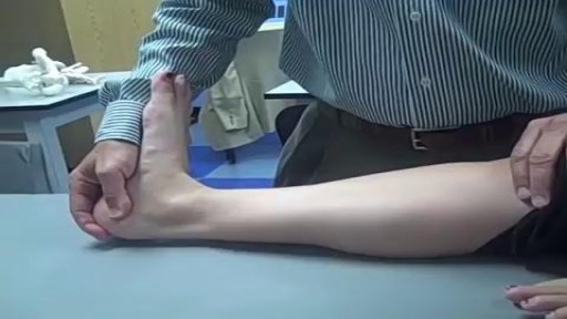
Homan’s sign test also called dorsiflexon sign test is a physical examination procedure that is used to test for Deep Vein Thrombosis (DVT). A positive Homan’s sign in the presence of other clinical signs may be a quick indicator of DVT. Clinical evaluation alone cannot be relied on for patient management, but when carefully performed, it remains useful in determining the need for additional testing (like D-dimer test, ultrasonography, multidetector helical computed axial tomography (CT), and pulmonary angiography) [1][2].

Minimally Invasive Surgery (MIS) Hip Joint Replacement is an advancement in hip replacement that offers important advantages over standard surgical procedures. Stryker has partnered with surgeons worldwide to develop MIS procedures and surgical instruments that are designed to help your surgeons do their very best to help you recover your lifestyle. These techniques bring together a wide variety of hip implants, new minimally invasive surgical techniques, and new instrumentation. The direct anterior approach is one of the minimally invasive techniques used in hip replacement surgery. Continuing orthopaedic experience suggests that this procedure may offer several advantages over the more traditional surgical approaches to hip replacement.1 Traditional hip replacement techniques involve operating from the side (lateral) or the back (posterior) of the hip, which requires a significant disturbance of the joint and connecting tissues and an incision approximately 8-12 inches long. In comparison, the direct anterior approach requires an incision that is only 3-4 inches in length and located at the front of the hip.1 In this position, the surgeon does not need to detach any of the muscles or tendons.

A very funny song made by the staff of the Ob/Gyn Gangnam style
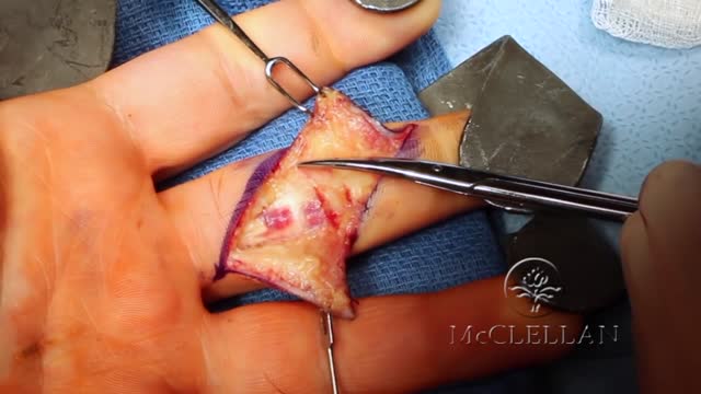
A deep cut on the palm side of your fingers, hand, wrist, or forearm can damage your flexor tendons, which are the tissues that help control movement in your hand. A flexor tendon injury can make it impossible to bend your fingers or thumb.
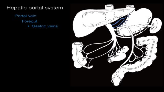
The hepatic portal system is the system of veins comprising the hepatic portal vein and its tributaries. It is responsible for directing blood from the region of the gastrointestinal tract between the esophagus and rectum and also includes venous drainage from the supplementary organs such as the spleen and pancreas.

A Bone scan or bone scintigraphy is a nuclear scanning test to find certain abnormalities in bone which are triggering the bone's attempts to heal. It is primarily used to help diagnose a number of conditions relating to bones, including: cancer of the bone or cancers that have spread (metastasized) to the bone, locating some sources of bone inflammation (e.g. bone pain such as lower back pain due to a fracture), the diagnosis of fractures that may not be visible in traditional X-ray images, and the detection of damage to bones due to certain infections and other problems.
Nuclear medicine bone scans are one of a number of methods of bone imaging, all of which are used to visually detect bone abnormalities. Such imaging studies include magnetic resonance imaging (MRI), X-ray computed tomography (CT) and in the case of 'bone scans' nuclear medicine. However, a nuclear bone scan is a functional test, which means it measures an aspect of bone metabolism, which most other imaging techniques cannot. The nuclear bone scan competes with the FDG-PET scan in seeing abnormal metabolism in bones, but it is considerably less expensive.
Nuclear bone scans are not to be confused with the completely different test often termed a "bone density scan," DEXA or DXA, which is a low exposure X-ray test measuring bone density to look for osteoporosis and other diseases where bones lose mass, without any bone re-building activity. The nuclear medicine scan technique is sensitive to areas of unusual bone re-building activity because the radiopharmaceutical is taken up by osteoblast cells which build bone. The technique therefore is sensitive to fractures and bone reaction to infections and bone tumors, including tumor metastases to bones, because all these pathologies trigger bone osteoblast activity. The bone scan is not sensitive to osteoporosis or multiple myeloma in bones, and therefore other techniques must be used to assess bone abnormalities from these diseases.
