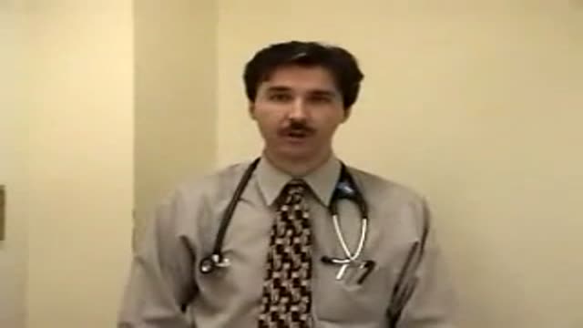
- Physical Examination
- Surgical Examination
- Ophthalmology
- Clinical Skills
- Orthopedics
- Surgery Videos
- Laparoscopy
- Pediatrics
- Funny Videos
- Cardiothoracic Surgery
- Nursing Videos
- Plastic Surgery
- Otorhinolaryngology
- Histology and Histopathology
- Neurosurgery
- Dermatology
- Pediatric Surgery
- Urology
- Dentistry
- Oncology and Cancers
- Anatomy Videos
- Health and Fitness
- Radiology
- Anaesthesia
- Physical Therapy
- Pharmacology
- Interventional Radiology
- Cardiology
- Endocrinology
- Gynecology
- Emergency Medicine
- Psychiatry and Psychology
- Childbirth Videos
- General Medical Videos
- Nephrology
- Physiology
- Diet and Food Health
- Diabetes Mellitus
- Neurology
- Women Health
- Osteoporosis
- Gastroenterology
- Pulmonology
- Hematology
- Rheumatology
- Toxicology
- Nuclear Medicine
- Infectious Diseases
- Vascular Disease
- Reproductive Health
- Burns and Wound Healing
- Other
Top videos
Pulmonary edema is usually caused by a heart condition. Other causes include pneumonia, exposure to certain toxins and drugs, and being at high elevations. Depending on the cause, pulmonary edema symptoms may appear suddenly or develop over time. Mild to extreme breathing difficulty can occur. Cough, chest pain, and fatigue are other symptoms. Treatment generally includes supplemental oxygen and medications.
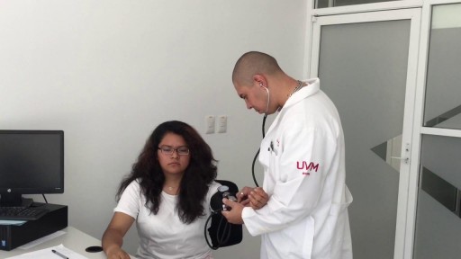
A detailed video showing how to clinically exam the abdomen
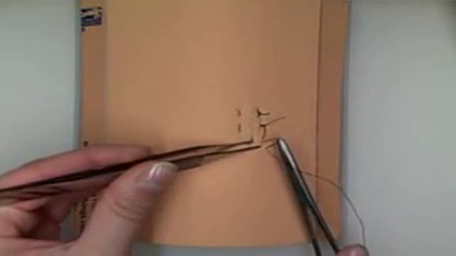
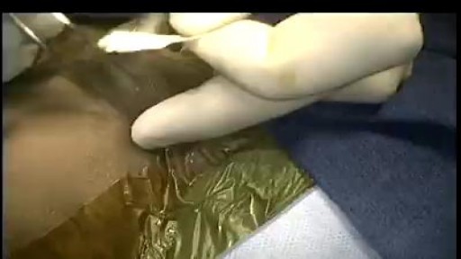
Demonstration of vertical mattress suturing technique for laceration repair or wound closure in the operating room.
Carotid Stenosis and what it means. The detection and treatment of carotid artery disease for the prevention of stroke is one of the most effective treatments in all of medicine.
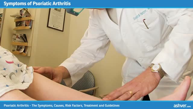
Psoriatic arthritis is a chronic arthritis. In some people, it is mild, with just occasional flare ups. In other people, it is continuous and can cause joint damage if it is not treated. Early diagnosis is important to avoid damage to joints. Psoriatic arthritis typically occurs in people with skin psoriasis, but it can occur in people without skin psoriasis, particularly in those who have relatives with psoriasis. Psoriatic arthritis typically affects the large joints, especially those of the lower extremities, distal joints of the fingers and toes, and also can affect the back and sacroiliac joints of the pelvis. For most people, appropriate treatments will relieve pain, protect the joints, and maintain mobility. Physical activity helps maintain joint movement. Psoriatic arthritis is sometimes misdiagnosed as gout, rheumatoid arthritis or osteoarthritis. - See more at: http://www.rheumatology.org/I-Am-A/Patient-Caregiver/Diseases-Conditions/Psoriatic-Arthritis#sthash.VsBTUw76.dpuf
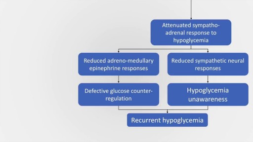
Hypoglycemia is a common and serious medical emergency which may occur in both daibetic and non-diabetic patients. The signs and symptoms of hypoglycaemia may be present in many individuals, but may also be masked in several individuals due to a condition called hypoglycaemia induced autonomic failure. This presentation aims to deal with the causes, clinical features, diagnosis and management of various causes of hypoglycaemia. The causes of hypoglycaemia may be divided into hypoglycaemia in ill or medicated individuals and hypoglycaemia in previously asymptomatic individuals. A variety of causes are discussed under both headings. Management of hypoglycaemia is also discussed in detail. There is also a brief discussion about management of insulinoma.
Minimally invasive surgery has been shown to be feasible and safe in pediatric patients since 1975 when laparoscopic surgery was first used to treat a small bowel obstruction. Laparoscopy is an option for surgical repair of inguinal hernias in addition to the traditional open approach.

Hemophagocytic lymphohistiocytosis is a rare but life threatening condition characterised by activation of macrophages which result in phagocytosis of RBCs and cytokine mediated tissue damage. This presentation aims to discuss the genetic basis, clinical features, diagnostic criteria and management options in this serious condition. The management options in HLH include Etoposide, Dexamethasone, Cyclosorine, Tacrolimus, Alemtuzumab and stem cell transplant.
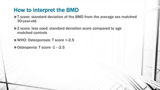
The discussion begins with a basic explanation of Bone biology taking into consideration the osteoblast and osteoclast balance. Concepts of RANK, RANK ligand and Osteoprotegerin are included. Risk factors for Osteoporosis such as Age, alcohol, smoking, sedentary lifestyle are also discussed.
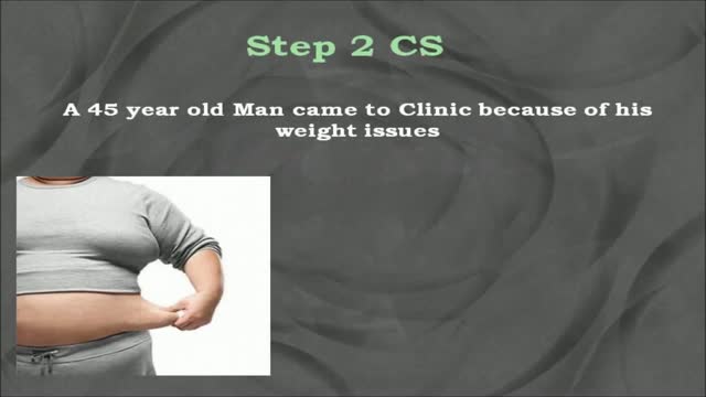
USMLE Step 2 CS - Obesity This is just preview video. To get full access please visit our website : www.usmletutoring.com

DOING LESS BUT BRAINY DESCRIBES A NEW GENERATION OF IMMEDIATE ZIRCONIA IMPLANTS ANATOMICAL AND CUSTOM-MADE. YOUR DENTAL ROOT IS MILLED IN ZIRCONIA AND IN 20 SECONDS SEATED, NO DRILLING, NO AUGMENTATION, NO MEMBRANES, FLAPLESS, NO 3D PLANNING, NO CAD/CAM SPLINTS OR GUIDED SURGERY REQUIRED! EASY AND CONSEQUENTIAL SYSTEM. NO MORE INCONGRUOUS AND UGLY SILVER-COLORED TITANIUM IMPLANTS IN TIME CONSUMING, PAINFUL AND COSTLY PROCEDURES. IT`S HIGH TIME TO RESPECT THE ANATOMY NOT ALTER IT BY DRILLING AND AUGMENTATION. BIOIMPLANT

UCLA Hand Transplant Procedure
Gonorrhea is a sexually transmitted disease (STD). It’s caused by infection with the bacterium Neisseria gonorrhoeae. It tends to infect warm, moist areas of the body, including the: urethra (the tube that drains urine from the urinary bladder) eyes throat vagina anus female reproductive tract (the fallopian tubes, cervix, and uterus) Gonorrhea passes from person to person through unprotected oral, anal, or vaginal sex. People with numerous sexual partners or those who don’t use a condom are at greatest risk of infection. The best protections against infection are abstinence, monogamy (sex with only one partner), and proper condom usage. Behaviors that make a person more likely to engage in unprotected sex also increase the likelihood of infection. These behaviors include alcohol abuse and illegal drug abuse, particularly intravenous drug use.
Systemic Lupus Erythematosus Animation 3d
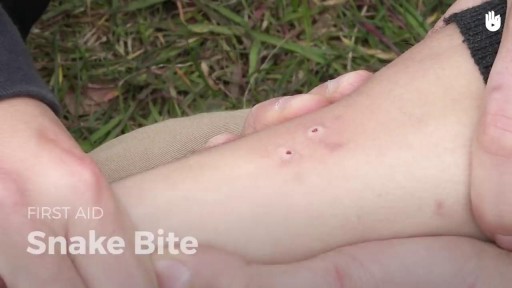
Move the person beyond striking distance of the snake. Have the person lie down with wound below the heart. Keep the person calm and at rest, remaining as still as possible to keep venom from spreading. Cover the wound with loose, sterile bandage
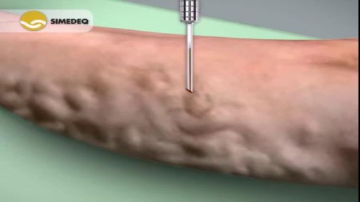
Varicose veins are generally benign. The cause of this condition is not known. For many people, there are no symptoms and varicose veins are simply a cosmetic concern. In some cases, they cause aching pain and discomfort or signal an underlying circulatory problem. Treatment involves compression stockings, exercise, or procedures to close or remove the veins.
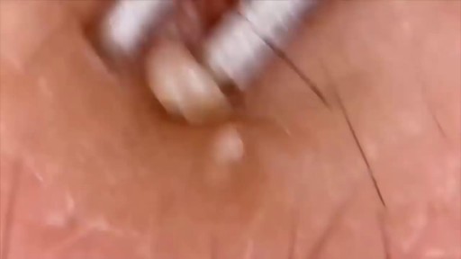
Watch that video of Popping Pimples
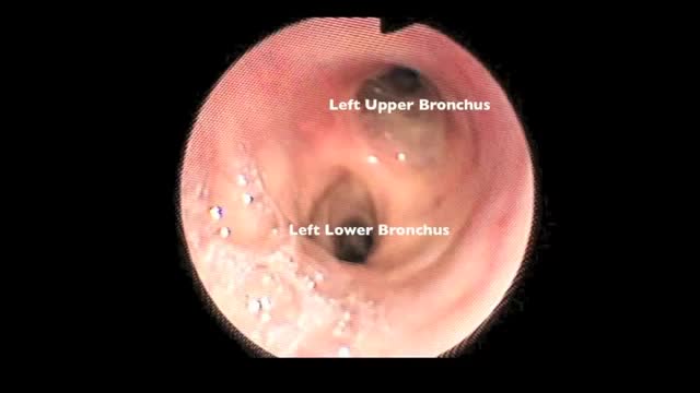
Flexible bronchoscopy is a procedure that allows a clinician to examine the breathing passages (airways) of the lungs (figure 1). Flexible bronchoscopy can be either a diagnostic procedure (to find out more about a possible problem) or a therapeutic procedure (to try to treat an existing problem or condition).
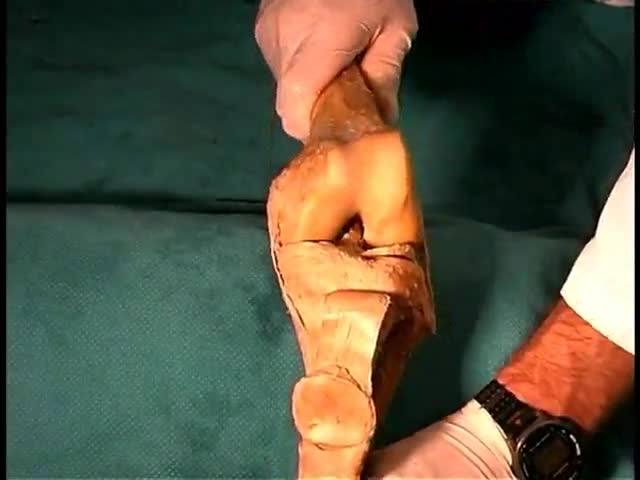
Anatomy of The Lower Limb Joints
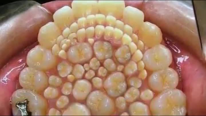
Watch that video of 232 Teeth Removal From Indians' Boy Mouth
