- Physical Examination
- Surgical Examination
- Ophthalmology
- Clinical Skills
- Orthopedics
- Surgery Videos
- Laparoscopy
- Pediatrics
- Funny Videos
- Cardiothoracic Surgery
- Nursing Videos
- Plastic Surgery
- Otorhinolaryngology
- Histology and Histopathology
- Neurosurgery
- Dermatology
- Pediatric Surgery
- Urology
- Dentistry
- Oncology and Cancers
- Anatomy Videos
- Health and Fitness
- Radiology
- Anaesthesia
- Physical Therapy
- Pharmacology
- Interventional Radiology
- Cardiology
- Endocrinology
- Gynecology
- Emergency Medicine
- Psychiatry and Psychology
- Childbirth Videos
- General Medical Videos
- Nephrology
- Physiology
- Diet and Food Health
- Diabetes Mellitus
- Neurology
- Women Health
- Osteoporosis
- Gastroenterology
- Pulmonology
- Hematology
- Rheumatology
- Toxicology
- Nuclear Medicine
- Infectious Diseases
- Vascular Disease
- Reproductive Health
- Burns and Wound Healing
- Other
Top videos
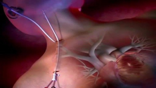
How a Clot Can Become a Pulmonary Embolism
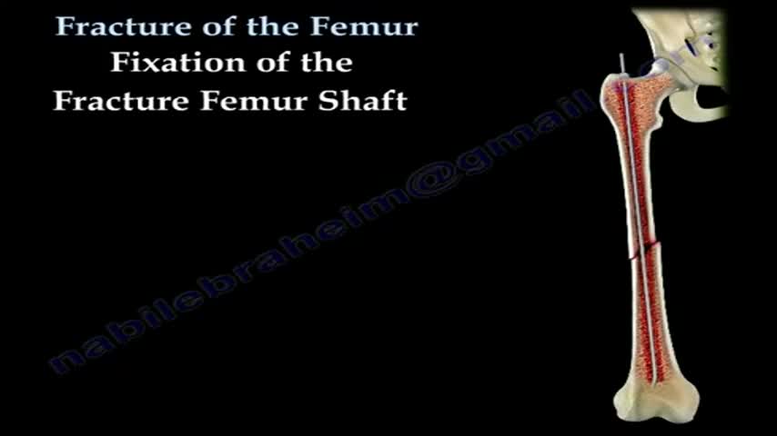
Open reduction and internal fixation (ORIF) is surgery used to stabilize and heal a broken bone. You might need this procedure to treat your broken thighbone (femur). The femur is the large bone in the upper part of your leg. Different kinds of trauma can damage this bone, causing it to fracture into 2 or more pieces. This might happen to the part of the femur near your knee, near the middle of the femur, or in the part of the femur that forms part of your hip joint. In certain types of femur fractures, your femur has broken, but its pieces still line up correctly. In other types of fractures (displaced fractures), the trauma moves the bone fragments out of alignment. If you fracture your femur, you usually need ORIF to bring your bones back into place and help them heal. During an open reduction, orthopedic surgeons reposition your bone pieces during surgery, so that they are back in their proper alignment. This contrasts with a closed reduction, in which a healthcare provider physically moves your bones back into place without surgically exposing your bone.
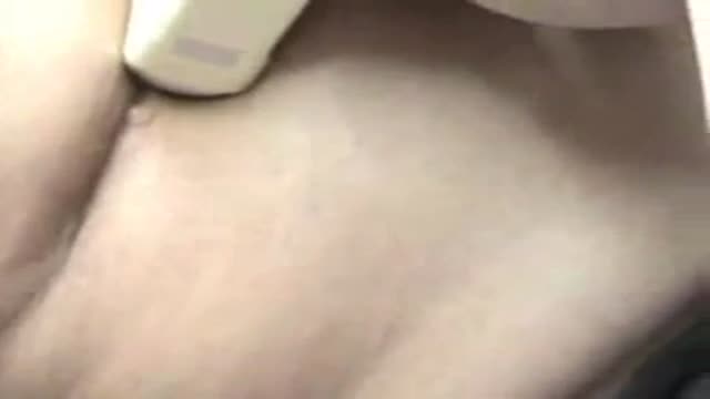
Patient 65-year-old of age who comes to the medical consultation with pain moderated pain in the right hypochondrium of “several years of evolution” but that it increased one week ago. Also, she shows pain in the umbilical region of “many years of evolution”, that is supported according to the patient - in a constant way.rnTo the examination, we observe an umbilical hernia, apparently divided into two parts. The hernia of the external region measures 25.1 centimeters x 18.0 centimeters and the one that occupies the average region measures 12.0 centimeters x 10.0 centimeters.rnPatient who comes to the medical consultation with moderated pain in the right hypochondrium of one year of evolution but it increased one week ago after eat duck.rnIn the ultrasound scan of the region of the right hypochondrium (patient came having breakfast, that is to say, without previous preparation ) we can observe the liver of 123.8 millimeters high, as well as the porta vein with a diameter of 7.3 millimeters.rnOn having observed the Gallbladder, we think that a side wall is increased in 2.7 mm (hyperechogenic) with several “echogenics points” in the interior (”Biliary Mud”).
The measurements of the gallbladder were: 39.0 x 17.4 millimeters.rnWe can appreciates an echogenic image in the interior that it would make think about stone. The stones are identified as echogenic foci casting acoustic shadowing but but this image did not appear and a re-evaluation is decided in 15 days.
Acalculous cholecystopathy which means disease or condition of the gallbladder without the presence of gallstones. You might also call it functional gallbladder disorder or impaired gallbladder emptying. Some causes may be chronic inflammation, a problem with the smooth muscles of the gallbladder or the muscle of the Sphincter of Oddi being too tight.
REMEMBER:
Umbilical hernia is a congenital malformation, especially common in infants of African descent, and more frequent in boys. An Acquired umbilical hernia directly results from increased intra-abdominal pressure and are most commonly seen in obese individuals.
Presentation:A hernia is present at the site of the umbilicus (commonly called a navel, or belly button) in the newborn; although sometimes quite large, these hernias tend to resolve without any treatment by around the age of 5 years. Obstruction and strangulation of the hernia is rare because the underlying defect in the abdominal wall is larger than in an inguinal hernia of the newborn. The size of the base of the herniated tissued is inversely correlated with risk of strangulation (i.e. narrow base is more likely to strangulate).
Babies are prone to this malformation because of the process during fetal development by which the abdominal organs form outside the abdominal cavity, later returning into it through an opening which will become the umbilicus.
Differential diagnosisrnImportantly this type of hernia must be distinguished from a paraumbilical hernia which occurs in adults and involves a defect in the midline near to the umbilicus, and from omphalocele.
Watch that video to know the Erectile Dysfunction - Cause and Treatment
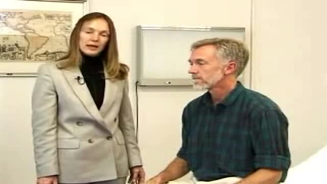
USMLE Step 2 CS - Shoulder Pain This is just preview video. To get full access please visit our website : www.usmletutoring.com
USMLE Step 2 CS - Weight loss This is just preview video. To get full access please visit our website : www.usmletutoring.com

Inguinal hernia repair without mesh, Desarda Repair, no recurrence, pain, no mesh hernia surgery, hernia operation, no mesh, without mesh, hernia operation, hernia surgery, new method.http://www.desarda.com
USMLE Step 2 CS - Numbness Weakness Full Video

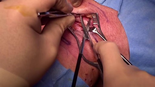
Dr. Nick demonstrates how to numb a toe for a patient who had a subungual hematoma “collection of blood under the nail”. This patient stubbed his toe and needed to have the nail removed.
#satisfying #reaction #amazing
MAKE SURE TO SUBSCRIBE FOR ALL THE NEW SURGICAL AND EDUCATIONAL VIDEOS COMING!!
👉🏻For more information visit :
https://drnickcampi.com
👉🏻Follow me on TikTok!!
https://vm.tiktok.com/ZMeXLbc5F/I’ll
👉🏻Connect with me!!
https://www.instagram.com/drnickcampitelli
👉🏻Check out this video of how we remove an ingrown toenail!
https://youtu.be/JyZo8aZDYds
👉🏻Dr. Nick Campitelli Performs latest Minimally Invasive Bunion Surgery! Watch this video!
https://youtu.be/eRpABMsCbOU
Dr. Nick Campitelli is a podiatrist who specializes in foot and ankle surgery in the Akron and Cleveland Ohio area. He is the Residency Director of the Western Reserve Hospital / University Hospital Podiatric Medicine and Surgery Residency Program.
*** All content found on the this YouTube video including: text, images, audio, or other formats were created for informational purposes only. The Content is not intended to be a substitute for professional medical advice, diagnosis, or treatment. Always seek the advice of your physician or other qualified health provider with any questions you may have regarding a medical condition. Never disregard professional medical advice or delay in seeking it because of something you heard on this video. ***
◦
Learn more about Merit Medical's ProGuide™ Chronic Dialysis
Yannas had been studying collagen, a protein found in human skin. Teaming up during the 1970s, the two made a polymer (a chemical compound made of multiple repeating units). Using collagen fibers and a long sugar molecule, they formed a porous (full of small holes) material resembling skin.
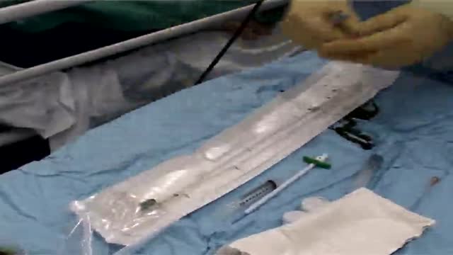
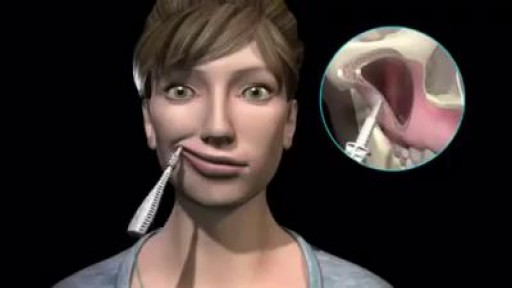
http://www.mediplus.co.uk A new and safer method of inserting a Foley catheter suprapubically. The technique allows the insertion to be carried out in an Outpatient setting, thus saving time, cost and effort. By using the Seldinger technique, the product reduces the chances of bowel or bladder perforation and resultant morbidity.
The product has been chosen by The NHS National Technology Adoption Centre to help facilitate adoption of the product.

Psychological testing refers to the administration of psychological tests. A psychological test is "an objective and standardized measure of a sample of behavior" (p. 4). The term sample of behavior refers to an individual's performance on tasks that have usually been prescribed beforehand.
Neurological Examination
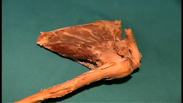
Anatomy of The Upper Limb Joints

Nearly 300 million people experience the world without certain colors every day. ‘Color For the Colorblind’ is a short documentary about what happened when we partnered with EnChroma, maker of color blindness-correcting glasses, to help people experience colors for the first time.
Sinus infections caused by viruses can use home (over-the-counter, OTC) treatments such as pain and fever medications (acetaminophen [Tylenol]), decongestants, and mucolytics. In addition, some health-care professionals suggest nasal irrigation or a sinus rinse solution to help relieve symptoms of sinus infections, even chronic sinusitis symptoms.
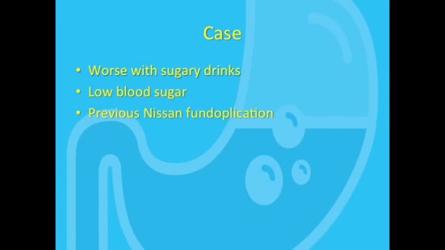
Dumping syndrome is a condition that can develop after surgery to remove all or part of your stomach or after surgery to bypass your stomach to help you lose weight. Also called rapid gastric emptying, dumping syndrome occurs when food, especially sugar, moves from your stomach into your small bowel too quickly.Diet: Eating too much sugar can cause sugars to pass into the colon, making the bacteria there get all excited and cause diarrhea. Other things like sorbitol, a sweetener in some sugarless candy, can also cause diarrhea through osmosis. Malabsorption: Some people don't digest sugars or fats properly.
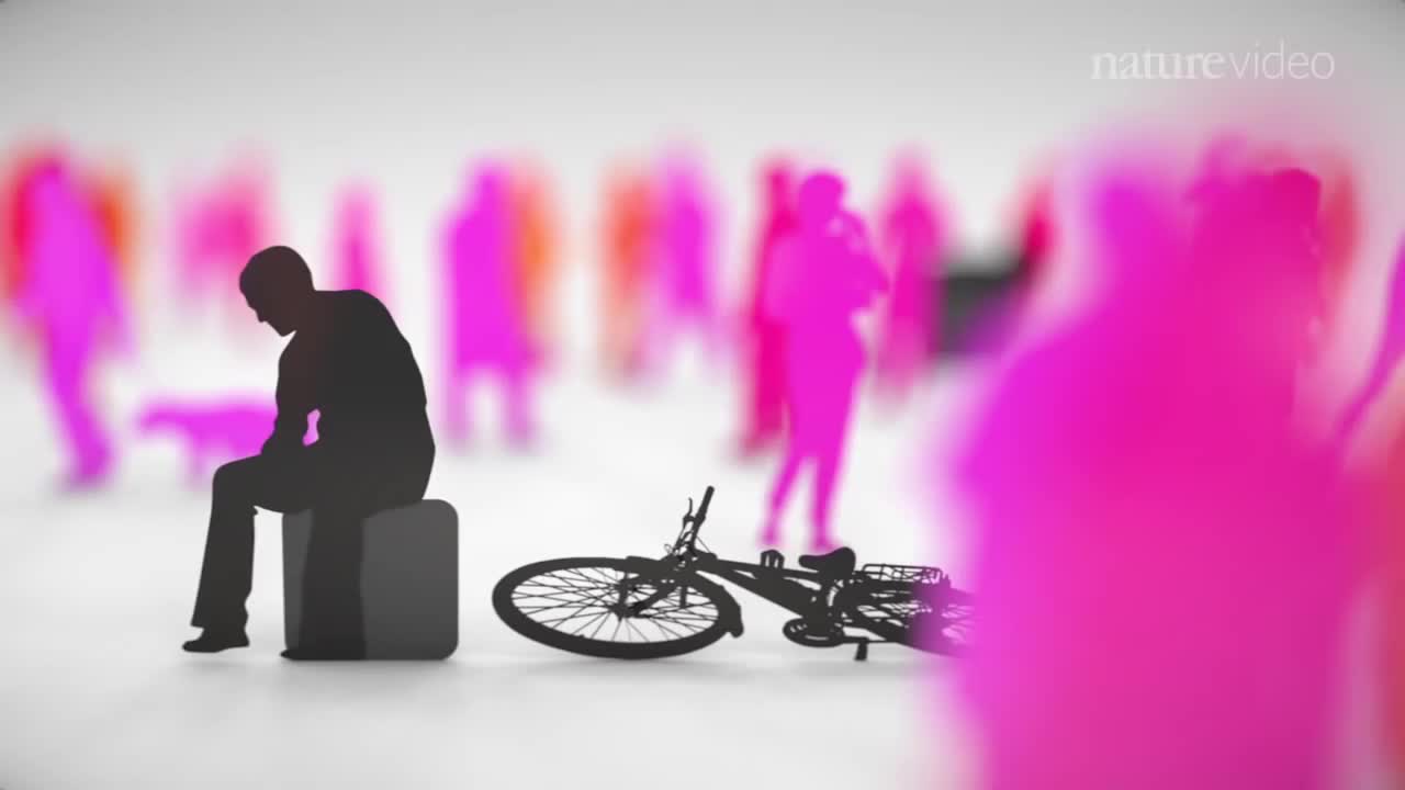
When you’re depressed, it can feel like you’ll never get out from under a dark shadow. However, even the most severe depression is treatable. So, if your depression is keeping you from living the life you want to, don’t hesitate to seek help. Learning about your depression treatment options will help you decide what approach is right for you. From therapy to medication to healthy lifestyle changes, there are many effective treatments that can help you overcome depression and reclaim your life.
