- Physical Examination
- Surgical Examination
- Ophthalmology
- Clinical Skills
- Orthopedics
- Surgery Videos
- Laparoscopy
- Pediatrics
- Funny Videos
- Cardiothoracic Surgery
- Nursing Videos
- Plastic Surgery
- Otorhinolaryngology
- Histology and Histopathology
- Neurosurgery
- Dermatology
- Pediatric Surgery
- Urology
- Dentistry
- Oncology and Cancers
- Anatomy Videos
- Health and Fitness
- Radiology
- Anaesthesia
- Physical Therapy
- Pharmacology
- Interventional Radiology
- Cardiology
- Endocrinology
- Gynecology
- Emergency Medicine
- Psychiatry and Psychology
- Childbirth Videos
- General Medical Videos
- Nephrology
- Physiology
- Diet and Food Health
- Diabetes Mellitus
- Neurology
- Women Health
- Osteoporosis
- Gastroenterology
- Pulmonology
- Hematology
- Rheumatology
- Toxicology
- Nuclear Medicine
- Infectious Diseases
- Vascular Disease
- Reproductive Health
- Burns and Wound Healing
- Other
Top videos

Force Does It Take To Break A Bone
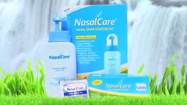
Are you seeking sinus, allergy, or nasal congestion relief? Nasal irrigation, also known as nasal rinsining, is your solution! Nasal Care's nasal irrigation system is an all-natural, simple, and easy sinus and allergy treatment that brings gentle and soothing sinus relief. Visit www.nasalcleanse.com to learn more about the safe, simple and all-natural relief you can experience with NasalCare's nasal irrigation system.
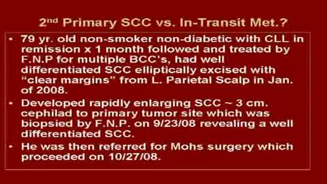
Graphic content of Mohs surgical removal of a large Squamous Cell Carcinoma on scalp followed by reconstruction with 10 week follow up. Visit us @ skincancercentre.com.
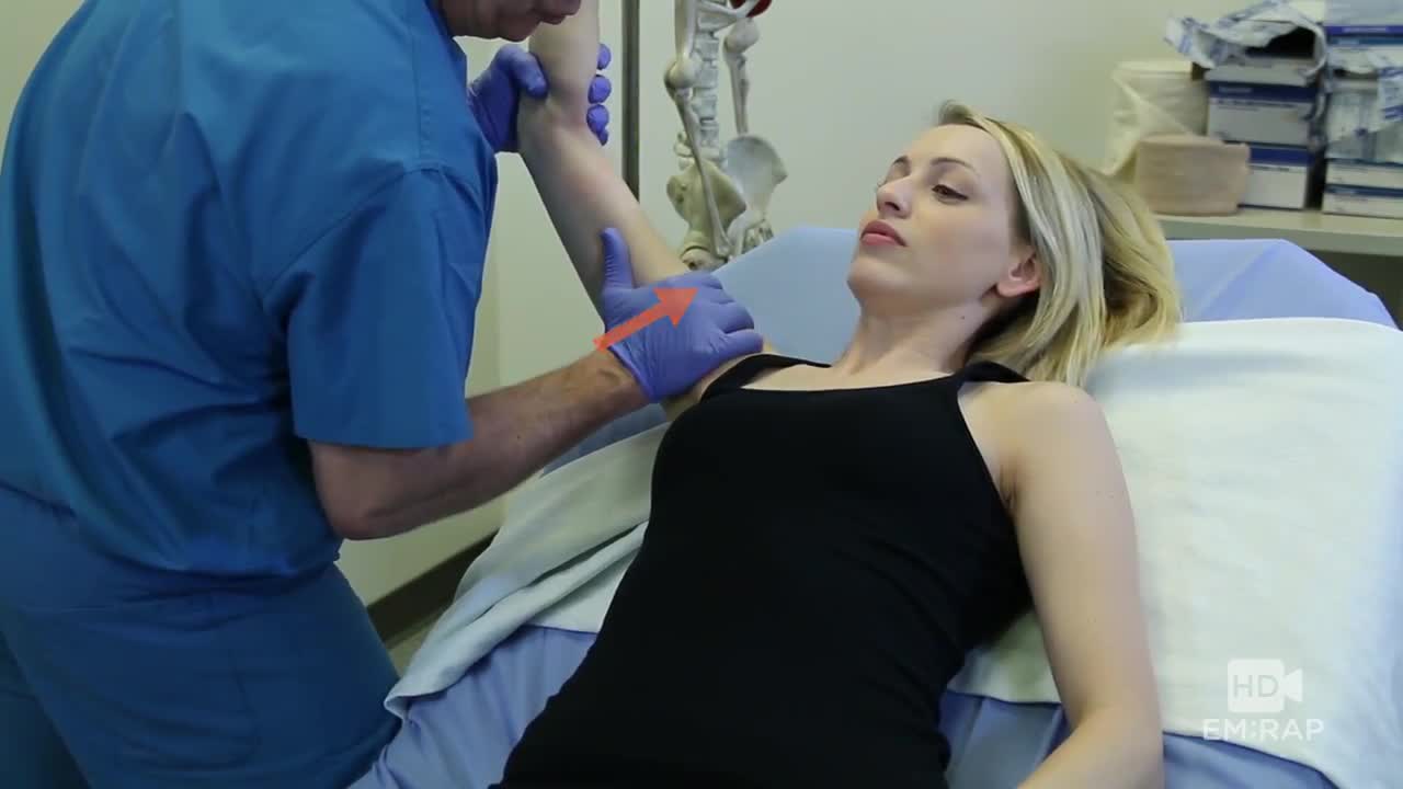
A technique for reducing an inferior shoulder dislocation. watch to learn more
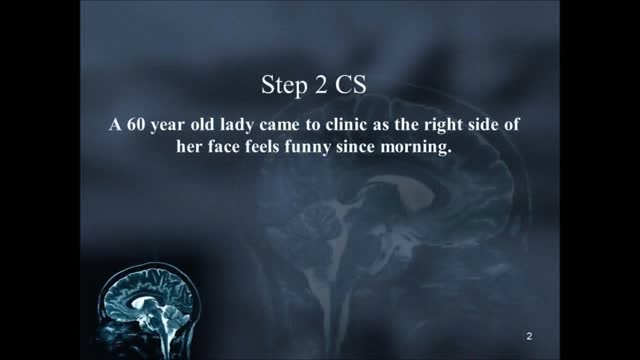
USMLE Step 2 CS - Numbness Weakness This is just preview video. To get full access please visit our website : www.usmletutoring.com
USMLE Step 2 CS - Vaginal Discharge This is just preview video. To get full access please visit our website : www.usmletutoring.com
USMLE Step 2 CS - Wrist Pain This is just preview video. To get full access please visit our website : www.usmletutoring.com

Preeclampsia is a pregnancy complication characterized by high blood pressure and signs of damage to another organ system, often the kidneys. Preeclampsia usually begins after 20 weeks of pregnancy in a woman whose blood pressure had been normal. Even a slight rise in blood pressure may be a sign of preeclampsia. Left untreated, preeclampsia can lead to serious — even fatal — complications for both you and your baby. If you have preeclampsia, the only cure is delivery of your baby. If you're diagnosed with preeclampsia too early in your pregnancy to deliver your baby, you and your doctor face a challenging task. Your baby needs more time to mature, but you need to avoid putting yourself or your baby at risk of serious complications.
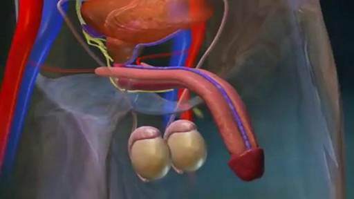
What Happens During an Erection?
In order to attain an erection, messages from the brain and other sense organs trigger the arteries of the penis to dilate. This allows an increased amount of blood to flow into three columns of spongy tissue in the penis.
As the arteries supplying blood to the corpus spongiosum and to the two larger columns, the corpus cavernosa, become filled with blood; the penis grows and becomes rigid. Pressure of the engorged tissue against the veins in the penis effectively traps blood within the penis until climax is reached or the sensation wanes.
What Are Penile Implants?
Impotence, or the inability to attain or maintain an erection, can be caused by a disruption at any stage in this process. Several types of penile implants are available that create an artificial erection. Two common types of implants are the semi-rigid malleable rod and the inflatable implant.
•The semirigid malleable rod is usually made of plastic with a core of flexible wire. These rods can be bent down to conceal the penis under clothing or raised to form an artificial erection.
•The inflatable implant is more complex and involves several working parts: a reservoir of fluid that is implanted into the abdomen, a pump system located in the scrotal sac near the testes, and two inflatable cylinders.
How Penile Implants Help Erectile Fuctioning
In order to attain an erection, the scrotal pump must be squeezed repeatedly to propel fluid into the penile cylinders. When an erection is no longer desired, a release valve is pressed on the side of the pump and the cylinders deflate.
Before Having Penile Implant Surgery
Persons considering these types of implants should speak with their physician or healthcare professional about possible risks and complications.
Lumbar puncture is a common emergency department procedure used to obtain information about the cerebrospinal fluid (CSF) for diagnostic and, less commonly, therapeutic reasons. Please refer to the full article on Lumbar Puncture for more details on the lumbar puncture procedure. Lumbar puncture is typically performed via “blind” surface landmark guidance. The surface landmark technique is reported to be successful in a high percentage of attempted lumbar punctures; however, surface landmark identification of underlying structures has been shown to be accurate only 30% of the time. [1] Unsuccessful identification of proper landmarks often leads to increased difficulty in obtaining CSF, if the procedure is performed, and a higher rate of complications. Few alternatives are available in these cases. If available, fluoroscopic-guided lumbar puncture may be performed. If not, treatment is sometimes initiated empirically without obtaining CSF. Disadvantages of using fluoroscopy include limited availability or necessary transport of the patient outside of the emergency department, inability to directly visualize the spinal canal, and inherent radiation exposure

Dr. Ankur Gupta of the Virginia Eye Institute discusses LASIK eye surgery as a method of correcting refractive errors. LASIK was first performed in Virginia on an FDA-approved laser by a VEI surgeon in 1996. Today, Virginia Eye Institute offers both conventional LASIK and custom LASIK with the bladeless IntraLase laser to precisely sculpt your cornea to correct refractive errors.
For more information on the services and procedures offered at Virginia Eye Institute please visit: https://goo.gl/6nX4RZ
THE CONTENT IN THIS VIDEO IS GENERAL IN NATURE AND DOES NOT SUBSTITUTE PROFESSIONAL MEDICAL ADVICE. The content on our website including, but not limited to, text, images, and videos is for informational and educational purposes only. Although we work hard to provide accurate general information, it is not a substitute for professional medical advice or consultations with healthcare professionals, and does not establish any kind of provider-patient relationship. Our website information is not intended to make any promises about the results of our products and services. We are not liable for actions taken based on content found on our website. If you are seeking medical advice, diagnoses, or treatment, we encourage you to call 804-287-2020 to make an appointment with one of our providers for your individualized care plan.
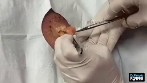
Glomus tumors are rare soft tissue neoplasms that typically present in adults (ages 20-40 years) as small, blue-red papules or nodules of the distal extremities, with most cases involving subungual sites. These tumors are typically painful, often causing paroxysmal pain in response to temperature changes or pressure. Glomus tumors are thought to arise from the glomus body, a thermoregulatory shunt concentrated in the fingers and toes. Most lesions are solitary and localized to cutaneous sites; however, generalized glomuvenous malformations, or multiple glomangiomas, have also been described, and may have extracutaneous involvement.

Dialysis lecture 1. Dialysis Study: EXPERT NOTES for DHA, Bonent, CHT, B.Sc in Dialysis, Diploma in Dialysis https://amzn.eu/d/35Ui1kT
2. Dialysis Study : Q & A: MCQs, Fill in the blanks, True or False https://amzn.eu/d/gGn8u73
1. Dialysis Study :EXPERT NOTES for DHA, Bonent, CHT, B.Sc in Dialysis, Diploma in Dialysis, Naseha Helal.
https://play.google.com/store/....books/details?id=D_7
2. Dialysis Study: Q & A MCQ https://play.google.com/store/....books/details?id=T_3
Whatsapp
https://chat.whatsapp.com/DKCHbgsNwXS1wd7xI31tpr
Telegram
https://t.me/dialysislife PRINCIPLE OF dialysis
https://youtu.be/cfOm0aFmbe8
Dialysis machine alarms
https://youtu.be/-1A1INyDEOg
DDS dialysis disequilibrium syndrome
https://youtu.be/8AqVFiBOkIc
Peritoneal Dialysis
https://youtu.be/iHPPadGmsv0
Itching
https://youtu.be/T83Wm3HHU4M
What is CRRT
https://youtu.be/jPgFnoSEBMU
LVH
https://youtu.be/ZhFL3Z6LHeA
Sorbent dialysis
https://youtu.be/-rie5dC_FkY
RO Water
https://youtu.be/3jlEsK4Lg_I
Carbon filter RO water
https://youtu.be/mJrgtjNafQw
Hemoperfusion
https://youtu.be/UkbBm8rm9Ww
AV fistula or Dialysis fistula
https://youtu.be/uDbyfqCkCbo
Dialysis MCQ
https://youtu.be/zmOj0BL6jVY
AVF cannulation
https://youtu.be/PyqMcHA07zY
Complications of AV fistula
https://youtu.be/a_CXIvuOO_s
Blood clotting during Dialysis
https://youtu.be/9hYNepiO2o8
Muscle crapms
https://youtu.be/09s07Eiqr2k
Hepatitis C
https://youtu.be/qdNj_GhmnSE
Dialysis procedure
https://youtu.be/u1mGqXO5pzQ
Hypotension
https://youtu.be/4EVPmWTSyN8
Heparin free dialysis
https://youtu.be/rFqAn7HcWwM
Plasmapheresis
https://youtu.be/kbgsjjs9krg
Isolated ultrafiltration
https://youtu.be/xp5I5--uWb0
High flux dialyzer
https://youtu.be/gCNsErn1HHM
Urea and Creatinine
https://youtu.be/Id9AIySMQ6c
Practical RO water demo
https://youtu.be/2pXKGMDNS84
Sodium profiling
https://youtu.be/bE_DcBXNB5g
Peritoneal Dialysis
https://youtu.be/vtK6VZsi8AY
Air embolism
https://youtu.be/WJE-xqnQfd8
Dialysate
https://youtu.be/z_nb43bcWsM
How to stop Bleed from fistula
https://youtu.be/N_inLKPhPUc
Dialysis short form
https://youtu.be/3BqB-gODb5o
Dialyzer reprocessing
https://youtu.be/XelfkKsndlc
Dialysis catheter
https://youtu.be/V7y90m4xlv8
How to set KT/V
https://youtu.be/hWXjU8VTQdk
Mircera injection
https://youtu.be/STtd3I3EijA
Dialysis procedure
https://youtu.be/MIdhIgcKRZ8
Dialysis in snake bite poison
https://youtu.be/niA9RI38jyY
Uf profiling
https://youtu.be/wyjpFjD5Hi0
Heparin dose
https://youtu.be/kB56MkzHIQ0
Hyperkalemia
https://youtu.be/1rWWNlcAuio
Change bandages of leaking fistula
https://youtu.be/_0cebWWdjM8
AvF needle
https://youtu.be/GvUxbXxftTk
Polycystic kidney disease
https://youtu.be/IhsMbHFXZG8
Nephrotic syndrome
https://youtu.be/FEEOsIrXxV8
Diabetic nephropathy
https://youtu.be/v-FBIQ7MA4k
Hemodialysis permanent access
https://youtu.be/_YrwxwiR0f8
Sex and dialysis
https://youtu.be/vvl8UT8lK4k
Albumin and dialysis
https://youtu.be/yzG7yD45Nwg
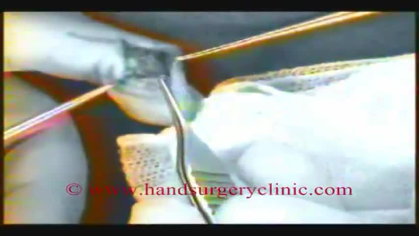
The procedure was performed under wrist block regional anesthesia with tourniquet control. A single Chinese finger trap was used on the thumb with 5 to 8 lb of ongitudinal traction. The arm was held down with wide tape around the tourniquet securing it to the hand table to serve as countertraction. A shoulder holder, rather than a traction tower, was used to facilitate fluoroscopic intervention more easily. The Trapeziometacarpal joint was detected by palpation. Joint distension was achieved by injecting 1 to 3 mL of normal saline (Fig. 1). It is important to distally direct the needle approximately 20 degrees to clear the dorsal flare of the metacarpal base and enter the joint capsule. This course should be reproduced upon entering with arthroscopic sleeve/ trocar assembly to minimize iatrogenic cartilage injury. Fluid distention is important to facilitate this. The incision for the 1-R (radial) portal, used for proper assessment of the dorsoradial ligament, posterior oblique ligament, and ulnar collateral ligament, was placed just volar to the abductor pollicis longus tendon. The incision for the 1-U (ulnar) portal, for better evaluation of the anterior oblique ligament and ulnar collateral ligament, was made just ulnar to the extensor pollicis brevis tendon. A short-barrel, 1.9-mm, 30- degree inclination arthroscope was used for complete visualization of the CMC joint surfaces, capsule, and ligaments, and then appropriate management was done, as dictated by the stage of the arthritis detected (Fig. 2A). A full-radius mechanical shaver with suction was used in all the cases, particularly for initial debridement and visualization. Most of the cases were augmented with radiofrequency ablation to perform a thorough synovectomy and radiofrequency was also used to perform chondroplasty in the cases with focal articular cartilage wear or fibrillation. Chondroplasty refers to thedebridement of the fibrillated cartilage to improve vascularity of the cartilage and enhance the growth of fibrocartilage. Ligamentous laxity and capsular attenu- ation were treated with thermal capsulorraphy using a radiofrequency shrinkage probe. We were careful to avoid thermal necrosis; hence, a striping technique was used to tighten the capsule of the lax joints. The striping technique refers to thermal shrinkage performed in longitudinal stripes on the lax capsule, so as to leave vascular zones between the stripes; hence, thermal necrosis is prevented. Arthroscopic stage I disease was characterized by synovitis without any cartilage wear, wherein a synovectomy coupled with thermal capsulor- raphy as described was performed.

Watch that Above Knee Leg Amputation Surgery
MASSIVE Goose Egg Drained
Heart Surgery | 8 Years Old Girl Open Heart Surgery Repair
A lipoma is a growth of fat cells in a thin, fibrous capsule usually found just below the skin. Lipomas aren't cancer and don't turn into cancer. They are found most often on the torso, neck, upper thighs, upper arms, and armpits, but they can occur almost anywhere in the body. One or more lipomas may be present at the same time.
Pharyngitis is caused by swelling in the back of the throat (pharynx) between the tonsils and the voice box (larynx). Most sore throats are caused by colds, the flu, coxsackie virus or mono (mononucleosis). Bacteria that can cause pharyngitis in some cases: Strep throat is caused by group A streptococcus.

What is Osteoporosis
