- Physical Examination
- Surgical Examination
- Ophthalmology
- Clinical Skills
- Orthopedics
- Surgery Videos
- Laparoscopy
- Pediatrics
- Funny Videos
- Cardiothoracic Surgery
- Nursing Videos
- Plastic Surgery
- Otorhinolaryngology
- Histology and Histopathology
- Neurosurgery
- Dermatology
- Pediatric Surgery
- Urology
- Dentistry
- Oncology and Cancers
- Anatomy Videos
- Health and Fitness
- Radiology
- Anaesthesia
- Physical Therapy
- Pharmacology
- Interventional Radiology
- Cardiology
- Endocrinology
- Gynecology
- Emergency Medicine
- Psychiatry and Psychology
- Childbirth Videos
- General Medical Videos
- Nephrology
- Physiology
- Diet and Food Health
- Diabetes Mellitus
- Neurology
- Women Health
- Osteoporosis
- Gastroenterology
- Pulmonology
- Hematology
- Rheumatology
- Toxicology
- Nuclear Medicine
- Infectious Diseases
- Vascular Disease
- Reproductive Health
- Burns and Wound Healing
- Other
Top videos
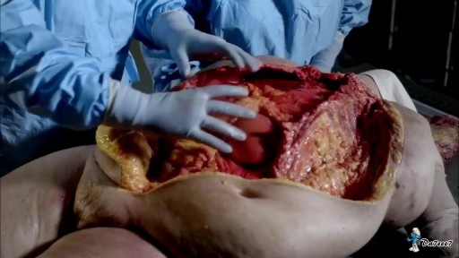
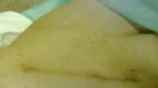
Watch that Cutting Inside Human Fat Body video

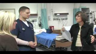
Children are special patients, and their medical needs are unique, including their surgical needs. At UNC Hospitals, an expert and experienced team of physicians treat children in a kid-friendly and family-centered environment. UNC Pediatric Surgeon Dr. Timothy Weiner explains

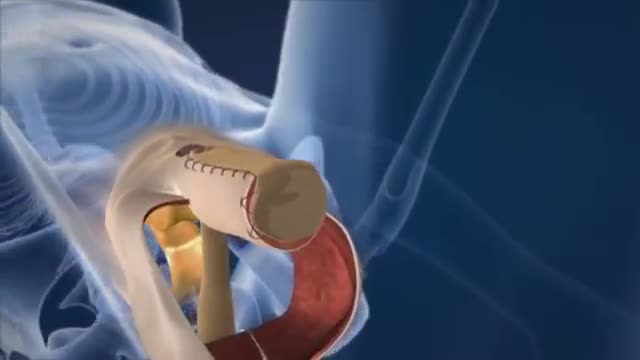
http://www.amerra.com In this patient education video from Colorectal Surgical Associates in Houston, Texas, learn more about the single incision laparoscopic colectomy procedure. This minimally invasive procedure uses a mini incision that
results in less pain, fewer complications, earlier recovery, and a smaller scar. Colorectal cancer is the second leading cause of cancer death in the United States. For more information please visit our website: www.csamd.com or call (713)-790-0600.
Watch that video of Virginity Hymen Repair Plastic Surgery

This video shows vaginal breech birth which is recommended to be delivered by C.Section in modern obstetrics
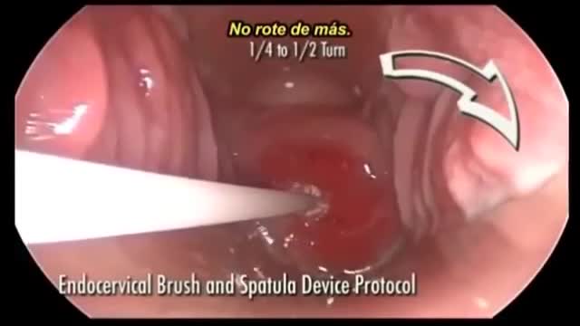
In patients age ;::25, HPV DNA testing is the preferred next step in management if the initial cytology shows ASC-US. In this method, samples are collected for both cytology and reflex HPV DNA. If cytology results are positive, HPV DNA testing is performed. If cytology results are negative, the sample for HPV DNA is discarded. HPV DNA testing along with Pap smear at 3 years is recommended if initial cytology shows ASC-US but HPV DNA testing is negative

a video showing the process of child birth or delivery using forceps
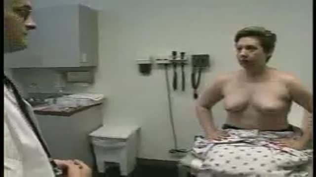
Full examination of the female from head to toe by Loyola Medical School, Chicago. Part 3
Watch that Male Catheter Insertion Procedure

It may be reassuring to know spotting or bleeding after sex is common and can come from the vagina, cervix, or urinary tract. It occurs most commonly in women 20 to 40 years old. Cervical Cancer: A very rare cause of spotting. ... Vaginal Dryness: Often caused by inadequate foreplay or vaginal lubrication.
Watch that video of Enema Medical Insertion Procedure
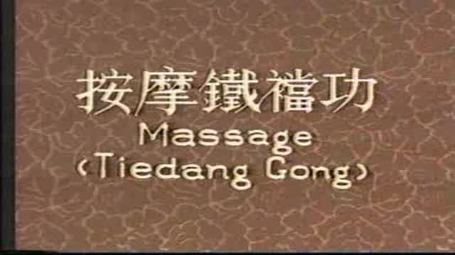
Mysterious massage from East Asia(CHINA).it can cure cure Erectile dysfunction,can let their life better.This video from mainland of China,so the language is Chinese mandarin.but you can see English show on the video too.Tiedang gong means kongfu of Iron penis&balls.
http://www.utexas.edu
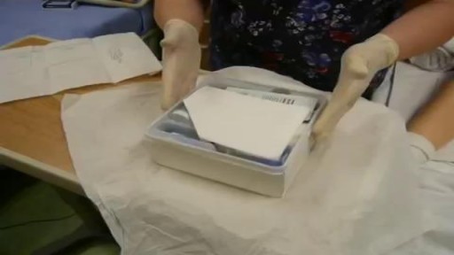
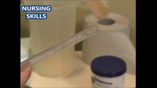
Nursing students practice their skills on mannequins and each other in the Nursing Skills Lab.
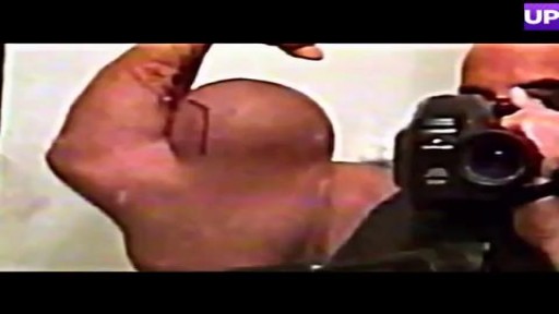
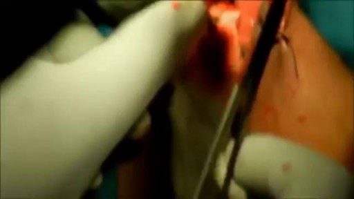
Bodybuilder Drains Synthol Hematoma From Bicep
A video showing the examination of femoral hernia.

Funny Video from hospital waiting room

It’s called gamma knife surgery, but there’s no cutting involved.
It’s been used at Mayo Clinic for 30 years as an alternative to open brain surgery.
The patient’s head is held still during the procedure with a headframe, which also serves as a map for the radiation. Using 3D imaging — typically an MRI — as a guide, the gamma knife is targeted directly at the tumor.
And with no hospital stay and minimal side effects, it’s a procedure that is efficient and can be lifesaving.
More health and medical news on the Mayo Clinic News Network. https://newsnetwork.mayoclinic.org/
Journalists: Clean and nat sound versions of this pkg available for download at https://newsnetwork.mayoclinic.org/
Register (free) at https://newsnetwork.mayoclinic.org/request-account/
Sex reassignment surgery for male-to-female involves reshaping the male genitals into a form with the appearance of, and, as far as possible, the function of female genitalia. Prior to any surgeries, patients usually undergo hormone replacement therapy (HRT), and, depending on the age at which HRT begins, facial hair removal. There are associated surgeries patients may elect to, including facial feminization surgery, breast augmentation, and various other procedures.
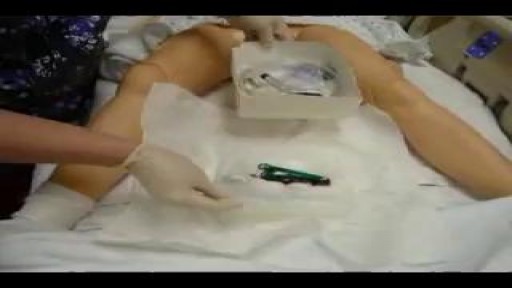
catheterization of the male urethra by a foley catheter
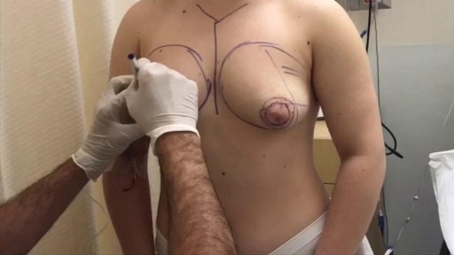
Tuberous breast deformity is a congenital breast anomaly that becomes manifest at the time of puberty and breast development. The three components of tubular deformity usually include, pseudoherniation of breast tissue into the nipple areolar complex, poorly defined inframammary fold and flattening of the lower pole of the breast which leads to a conical tubular shape. Stuart Linder M.D. 9675 BRIGHTON WAY, SUITE 420 BEVERLY HILLS CA 90210 (310) 275-4513
