- Physical Examination
- Surgical Examination
- Ophthalmology
- Clinical Skills
- Orthopedics
- Surgery Videos
- Laparoscopy
- Pediatrics
- Funny Videos
- Cardiothoracic Surgery
- Nursing Videos
- Plastic Surgery
- Otorhinolaryngology
- Histology and Histopathology
- Neurosurgery
- Dermatology
- Pediatric Surgery
- Urology
- Dentistry
- Oncology and Cancers
- Anatomy Videos
- Health and Fitness
- Radiology
- Anaesthesia
- Physical Therapy
- Pharmacology
- Interventional Radiology
- Cardiology
- Endocrinology
- Gynecology
- Emergency Medicine
- Psychiatry and Psychology
- Childbirth Videos
- General Medical Videos
- Nephrology
- Physiology
- Diet and Food Health
- Diabetes Mellitus
- Neurology
- Women Health
- Osteoporosis
- Gastroenterology
- Pulmonology
- Hematology
- Rheumatology
- Toxicology
- Nuclear Medicine
- Infectious Diseases
- Vascular Disease
- Reproductive Health
- Burns and Wound Healing
- Other
Top videos

In this medical video: This 72-year-old patient was unable to resist blinking when we tapped on the glabella. This is the glabellar reflex or Myerson's sign . It is often an early sign of Parkinson's disease, but can also be seen in early dementia as well as other progressive neurologic illness. Note the left (i.e., asymmetrical) hand resting tremor.
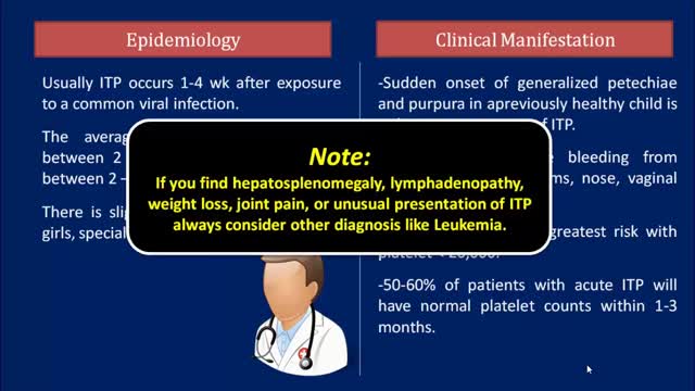
Idiopathic thrombocytopenic purpura (ITP) is a disorder that can lead to easy or excessive bruising and bleeding. The bleeding results from unusually low levels of platelets — the cells that help blood clot. Idiopathic thrombocytopenic purpura, which is also called immune thrombocytopenia, affects children and adults. Children often develop ITP after a viral infection and usually recover fully without treatment. In adults, the disorder is often long term. If you don't have signs of bleeding and your platelet count isn't too low, you may not need any treatment. In rare cases, the number of platelets may be so low that dangerous internal bleeding occurs. Treatment options are available.
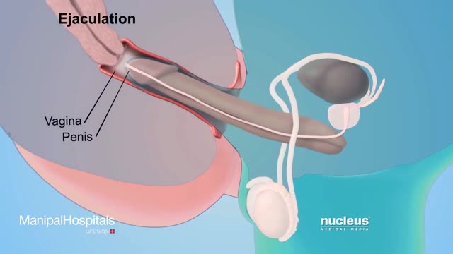
How To Use Male Condom Correctly - Manipal Hospitals
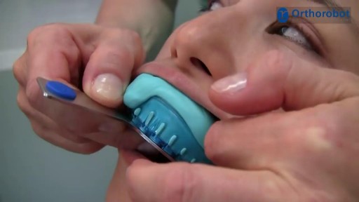
A short introduction on how to take a correction impression. The shown materials are recommended by Orthorobot and have proven to be fully compatible with the Orthorobot lab procedure.
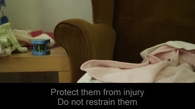
Pediatric febrile seizures, which represent the most common childhood seizure disorder, exist only in association with an elevated temperature. Evidence suggests, however, that they have little connection with cognitive function, so the prognosis for normal neurologic function is excellent in children with febrile seizures. [1] Epidemiologic studies have led to the division of febrile seizures into 3 groups, as follows: Simple febrile seizures Complex febrile seizures Symptomatic febrile seizures Essential update: Starting MMR/MMRV vaccination earlier may reduce seizure risk In a case-series analysis of a cohort of 323,247 US children born from 2004 to 2008, Hambidge et al found that delaying the first dose of measles-mumps-rubella (MMR) or measles-mumps-rubella-varicella (MMRV) vaccine beyond the age of 15 months may more than double the risk of postvaccination seizures in the second year of life. [2, 3] In infants, there was no association between vaccination timing and postvaccination seizures. [3] In the second year of life, however, the incident rate ratio (IRR) for seizures within 7-10 days was 2.65 (95% confidence interval [CI], 1.99-3.55) after first MMR doses at 12-15 months of age, compared with 6.53 (95% CI, 3.15-13.53) after first MMR doses at 16-23 months. For the MMRV vaccine, the IRR for seizures was 4.95 (95% CI, 3.68-6.66) after first doses at 12-15 months, compared with 9.80 (95% CI, 4.35-22.06) for first doses at 16-23 months.
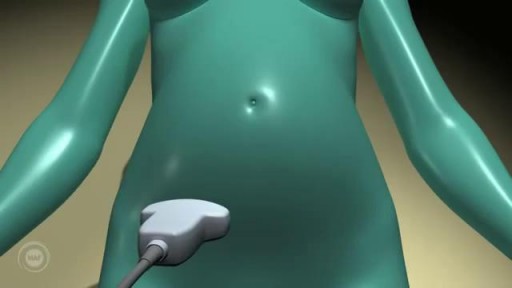
A prenatal ultrasound (also called a sonogram) is a noninvasive diagnostic test that uses sound waves to create a visual image of your baby, placenta, and uterus, as well as other pelvic organs. It allows your healthcare practitioner to gather valuable information about the progress of your pregnancy and your baby's health. During the test, an ultrasound technician (sonographer) transmits high-frequency sound waves through your uterus that bounce off your baby. A computer then translates the echoing sounds into video images that reveal your baby's shape, position, and movements. (Ultrasound waves are also used in the handheld instrument called a Doppler that your practitioner uses during your prenatal visits to listen to your baby's heartbeat.) You may have an early ultrasound at your practitioner's office at 6 to 10 weeks to confirm and date the pregnancy. Or you may not have one until the standard midpregnancy ultrasound between 16 and 20 weeks. That's when you may learn your baby's sex, if you like. (The technician will probably present you with a grainy printout of the sonogram as a keepsake.) You may also have a sonogram as part of a genetic test, such as the nuchal translucency test, chorionic villus sampling, or amniocentesis, or at any other time if there are signs of a problem with your baby. You'll have more frequent ultrasounds if you have diabetes, hypertension, or other medical complications.
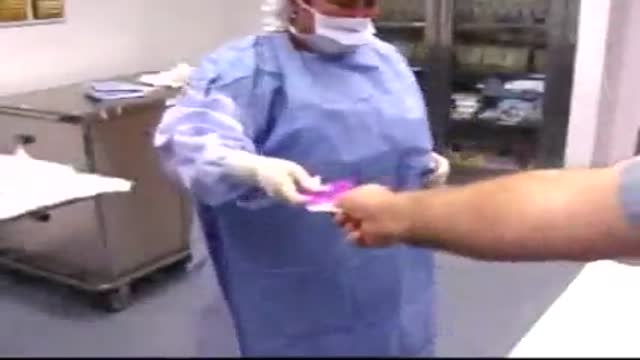
Turning To Seal Gown
Laparoscopic Uterosacral Colpoplexy HD
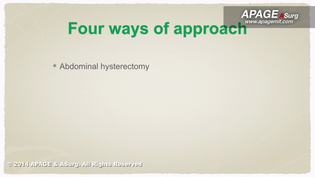
procedure is usually done in the hospital or outpatient surgical center under general anesthesia (while you are asleep and pain-free). The procedure is performed in the following way: The surgeon makes a small cut (incision) below the belly button (navel). A needle or tube is inserted into the incision. Carbon dioxide gas is passed into the abdomen through the needle or tube. The gas helps expand the area, giving the surgeon more room to work, and helping the surgeon see the organs more clearly. A tube is placed through the cut in your abdomen. A tiny video camera (laparoscope) goes through this tube and is used to see the inside of your pelvis and abdomen. More small cuts may be made if other instruments are needed to get a better view of certain organs. If you are having gynecologic laparoscopy, dye may be injected into your cervix area so the surgeon can view your fallopian tubes. After the exam, the gas, laparoscope, and instruments are removed, and the cuts are closed. You will have bandages over those areas.
![So You Want to Be a CARDIOTHORACIC SURGEON [Ep. 13]](https://i.ytimg.com/vi/sdxz242qDFA/maxresdefault.jpg)
So you want to be a cardiothoracic surgeon. You like the idea of open heart surgery and the glory that comes with being a CT surgeon. Let’s debunk the public perception myths of what it means to be a cardiothoracic surgeon, and give it to you straight. This is the reality of cardiothoracic surgery.
✒️ Accompanying Blog Post: https://medschoolinsiders.com/....medical-student/so-y
💌 Sign up for my weekly newsletter - https://medschoolinsiders.com/newsletter
🌍 Website & blog - https://medschoolinsiders.com
📸 Instagram - https://instagram.com/medschoolinsiders
🐦 Twitter - https://twitter.com/medinsiders
🗣️ Facebook - https://facebook.com/medschoolinsiders
🎥 My Youtube Gear: https://kit.co/kevinjubbalmd/
👀 Hand Picked Productivity Tools: https://www.amazon.com/shop/medschoolinsiders
🎵My Study Playlist: https://open.spotify.com/user/....1231934998/playlist/
TIME STAMPS:
00:41 - What is Cardiothoracic Surgery?
04:08 - How to Become a Cardiothoracic Surgeon
06:29 - Subspecialties within Cardiothoracic Surgery
07:49 - What You’ll Love About Cardiothoracic Surgery
09:10 - What You Won’t Love About Cardiothoracic Surgery
10:04 - Should You Become a Cardiothoracic Surgeon?
LINKS FROM VIDEO:
So You Want to Be Playlist: https://www.youtube.com/playli....st?list=PL2ADAFpTg5a
Day in the Life Playlist: https://www.youtube.com/playli....st?list=PLTCN43UFAlB
#medicalschool #cardiothoracicsurgery #soyouwanttobe
====================
Disclaimer: Content of this video is my opinion and does not constitute medical advice. The content and associated links provide general information for general educational purposes only. Use of this information is strictly at your own risk. Kevin Jubbal, M.D. and Med School Insiders LLC will not assume any liability for direct or indirect losses or damages that may result from the use of information contained in this video including but not limited to economic loss, injury, illness or death. May include affiliate links to Amazon. As an Amazon Associate, I may earn a commission on qualifying purchases made through them (at no extra cost to you).
How do you make a working human heart? Scientists can turn stem cells into beating heart cells, but getting them to organize into a 3D heart requires a scaffold. At the Massachusetts General Hospital in Boston, Harald Ott and his team are reusing the scaffold that nature provides. They’re stripping away all the living cells from dead hearts, before filling in the leftover matrix with healthy new cells. In this video, Brendan Maher finds out how the technique could be used to develop parts of the heart, like the aortic root and valve, for transplant.

A very simplified method giving information about cystic fibrosis

Watch that video of an Indian Boy Was Born With 232 Teeth Got Them Removed
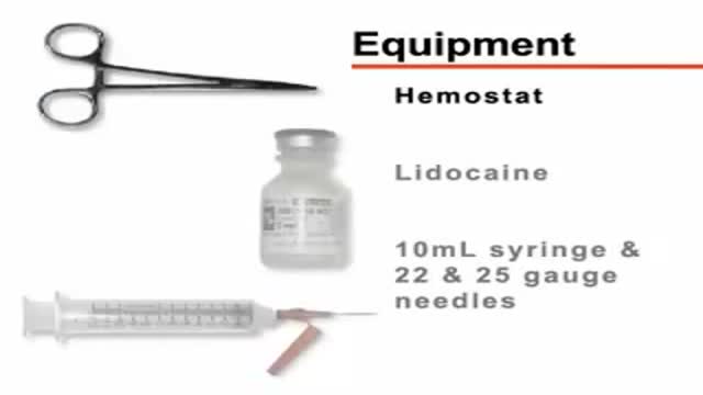
Thoracentesis is a procedure to remove fluid or air from around the lungs. A needle is put through the chest wall into the pleural space. The pleural space is the thin gap between the pleura. The pleura are a double layer of membranes that surrounds the lungs.
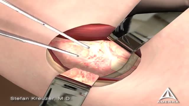
Femoroacetabular impingement (FAI) is a condition in which extra bone grows along one or both of the bones that form the hip joint — giving the bones an irregular shape. Because they do not fit together perfectly, the bones rub against each other during movement. Over time this friction can damage the joint, causing pain and limiting activity.
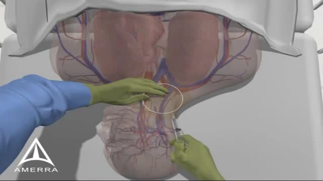
Catheters can be placed in veins in the neck (internal jugular vein), chest (subclavian vein or axillary vein), groin (femoral vein), or through veins in the arms (also known as a PICC line, or peripherally inserted central catheters).

Eric knew he needed help when an old knee injury began worsening over the course of time and was significantly affecting his quality of life. That’s when he turned to his hometown orthopedic experts at Mayo Clinic Health System in Mankato, who recommended a total knee replacement. After overcoming some initial fears, Eric decided it was time to have the operation — a fuller and more active life with his family depended on it.
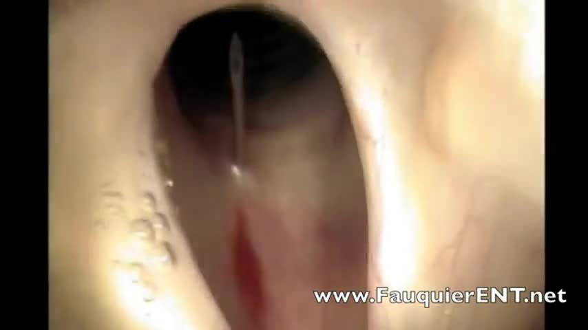
This video demonstrates how bronchoscopy and vocal cord mass injections can be performed under endoscopic guidance in a patient without any sedation. Only topical and local anesthesia is used for patient comfort.
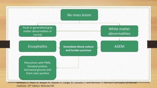
A detailed discussion of the causes, diagnosis and management of the causes of Meningitis and Encephalitis. Includes bacterial, viral, fungal and autoimmune conditions as well as treatment of these conditions. Includes antivirals such as Aciclovir and Ganciclovir as well as IVIG and plasma exchange for autoimmune encephalitis.
To get started, you need to find your pelvic floor muscles by stopping urination in midstream. If you succeed, you have located the right muscles. Once you have located your pelvic floor muscles, tighten the contraction for about 5 seconds, before relaxing for another 5 seconds.
