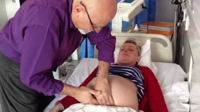
- Physical Examination
- Surgical Examination
- Ophthalmology
- Clinical Skills
- Orthopedics
- Surgery Videos
- Laparoscopy
- Pediatrics
- Funny Videos
- Cardiothoracic Surgery
- Nursing Videos
- Plastic Surgery
- Otorhinolaryngology
- Histology and Histopathology
- Neurosurgery
- Dermatology
- Pediatric Surgery
- Urology
- Dentistry
- Oncology and Cancers
- Anatomy Videos
- Health and Fitness
- Radiology
- Anaesthesia
- Physical Therapy
- Pharmacology
- Interventional Radiology
- Cardiology
- Endocrinology
- Gynecology
- Emergency Medicine
- Psychiatry and Psychology
- Childbirth Videos
- General Medical Videos
- Nephrology
- Physiology
- Diet and Food Health
- Diabetes Mellitus
- Neurology
- Women Health
- Osteoporosis
- Gastroenterology
- Pulmonology
- Hematology
- Rheumatology
- Toxicology
- Nuclear Medicine
- Infectious Diseases
- Vascular Disease
- Reproductive Health
- Burns and Wound Healing
- Other
Top videos
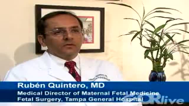
Highlights of a fetal laser surgery for twin-to-twin transfusion syndrome (TTTS) will be shown from Tampa General Hospital
TTTS affects 10 to 15 percent of identical-twin pregnancies and is the result of abnormal blood exchange between identical twins through a common placenta. The larger of the twins, or recipient, is surrounded by too much amniotic fluid and in danger of heart failure as its body tries to pump the overwhelming volume of blood intended for both. The smaller, or donor twin, is encased in a shrinking amniotic sac deprived of blood. Without treatment, both will likely die.
Rubén Quintero, M.D., Medical Director of Maternal Fetal Medicine/Fetal Surgery at Tampa General Hospital and Professor and Director of the Division of Maternal Fetal Medicine, Department of Obstetrics and Gynecology, University of South Florida College of Medicine will narrate the procedure and answer e-mail questions live as taped highlights of the procedure are shown.
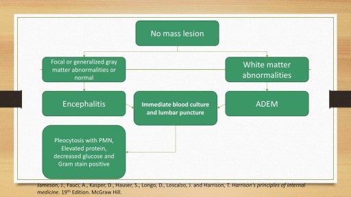
A detailed discussion of the causes, diagnosis and management of the causes of Meningitis and Encephalitis. Includes bacterial, viral, fungal and autoimmune conditions as well as treatment of these conditions. Includes antivirals such as Aciclovir and Ganciclovir as well as IVIG and plasma exchange for autoimmune encephalitis.

Shoulder Injection

We are aware that the "official" way to use an ear candle is small end down into the ear, but for this video, we have elected to use it the way most "lay" public would (small end up). Ear candling is an alternative medicine practice that is thought to remove earwax. However, this video illustrates how ineffective this practice is in removing earwax... and can potentially be even harmful. And yes... It is still frequently practiced.
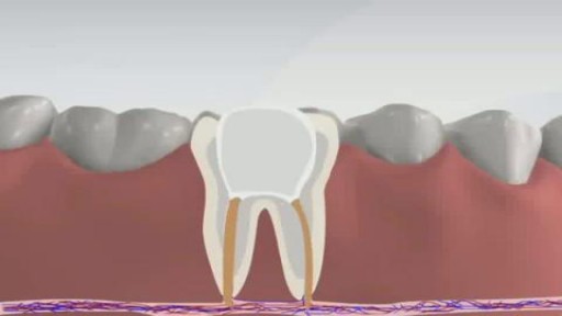
Root canal is a treatment to repair and save a badly damaged or infected tooth instead of removing it. The term "root canal" comes from cleaning of the canals inside a tooth's root. Decades ago, root canal treatments often were painful. With dental advances and local anesthetics, most people have little if any pain with a root canal. In fact, it's probably more painful living with a decayed tooth. Root canal alternatives include extracting the damaged tooth and replacing it with a dental implant, bridge or removable partial denture.
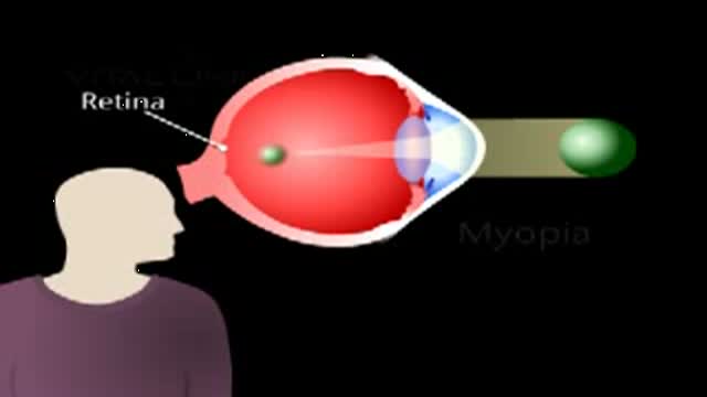
This animated video reviews myopia, which is the medical term for nearsightedness.
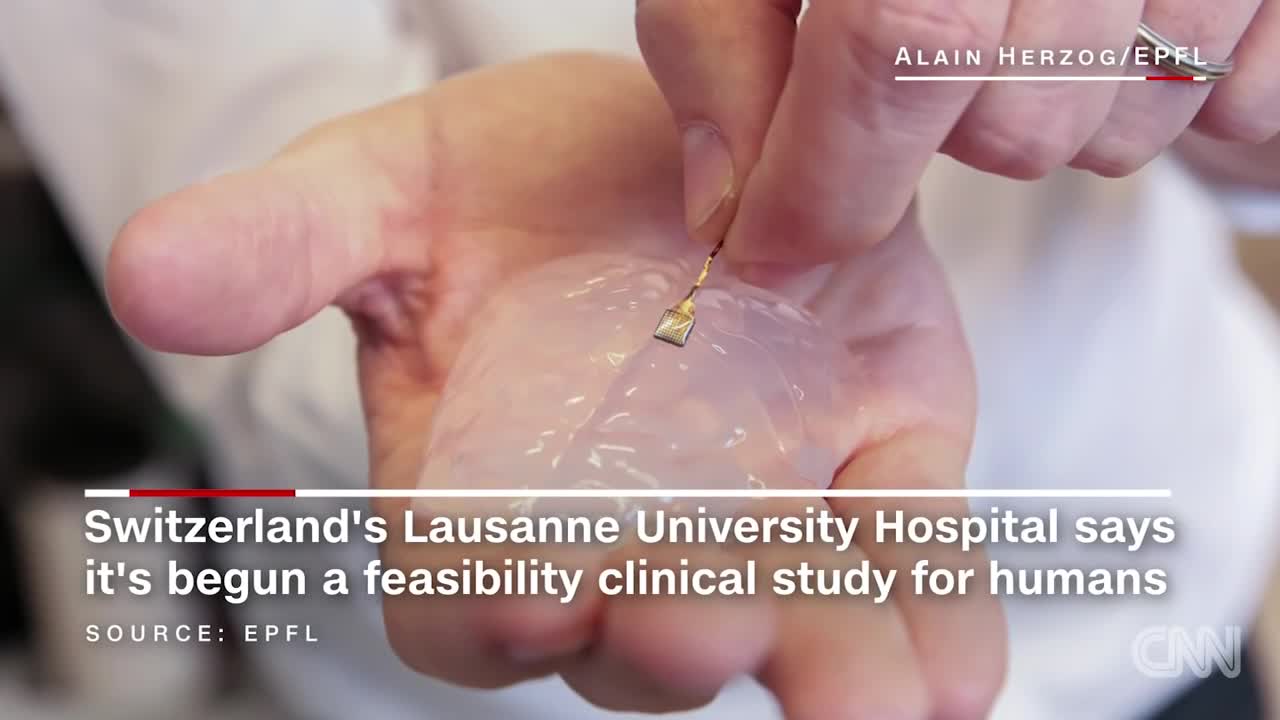
Scientists have developed a wireless brain implant that enabled a paralyzed monkey to walk again.
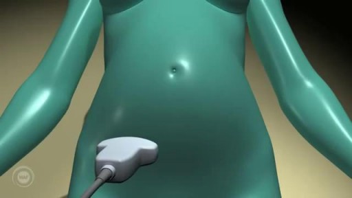
A prenatal ultrasound (also called a sonogram) is a noninvasive diagnostic test that uses sound waves to create a visual image of your baby, placenta, and uterus, as well as other pelvic organs. It allows your healthcare practitioner to gather valuable information about the progress of your pregnancy and your baby's health. During the test, an ultrasound technician (sonographer) transmits high-frequency sound waves through your uterus that bounce off your baby. A computer then translates the echoing sounds into video images that reveal your baby's shape, position, and movements. (Ultrasound waves are also used in the handheld instrument called a Doppler that your practitioner uses during your prenatal visits to listen to your baby's heartbeat.) You may have an early ultrasound at your practitioner's office at 6 to 10 weeks to confirm and date the pregnancy. Or you may not have one until the standard midpregnancy ultrasound between 16 and 20 weeks. That's when you may learn your baby's sex, if you like. (The technician will probably present you with a grainy printout of the sonogram as a keepsake.) You may also have a sonogram as part of a genetic test, such as the nuchal translucency test, chorionic villus sampling, or amniocentesis, or at any other time if there are signs of a problem with your baby. You'll have more frequent ultrasounds if you have diabetes, hypertension, or other medical complications.
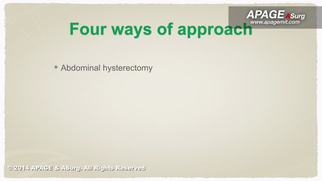
procedure is usually done in the hospital or outpatient surgical center under general anesthesia (while you are asleep and pain-free). The procedure is performed in the following way: The surgeon makes a small cut (incision) below the belly button (navel). A needle or tube is inserted into the incision. Carbon dioxide gas is passed into the abdomen through the needle or tube. The gas helps expand the area, giving the surgeon more room to work, and helping the surgeon see the organs more clearly. A tube is placed through the cut in your abdomen. A tiny video camera (laparoscope) goes through this tube and is used to see the inside of your pelvis and abdomen. More small cuts may be made if other instruments are needed to get a better view of certain organs. If you are having gynecologic laparoscopy, dye may be injected into your cervix area so the surgeon can view your fallopian tubes. After the exam, the gas, laparoscope, and instruments are removed, and the cuts are closed. You will have bandages over those areas.

Neglected elbow dislocations are seen in patients hailing from Africa and Asia. A Nigerian patient with this condition was successfully treated by open reduction and external fixator application

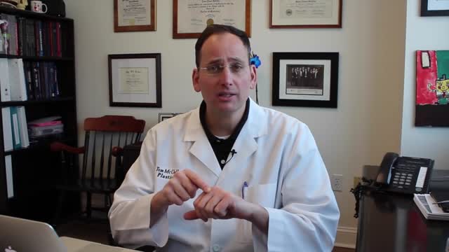
An Abdominoplasty (commonly referred to as a “Tummy Tuck”) removes excess fat and skin around your abdomen to shape and contour your midsection. During surgery, I also restore weakened or separated muscles to help create an abdominal profile that is both; smoother and more firm.
Watch this video as we go from the operating table to her 2-month post-op results!
If you’re interested in learning more about tummy tuck surgery or any other services we offer, please DM us or give us a call today!
☎️(424) 266-4181
🌐DrJohnDiaz.com
#DrJohnDiaz #DrDiaz #BeverlyHills #BeverlyHillsPlasticSurgery #BeverlyHillsPlasticSurgeon #DiazPlasticSurgery #PlasticSurgery #PlasticSurgeon #TummyTuck #Abdominoplasty #BeverlyHillsTummyTuck #TummyTuckBeverlyHills #AbdominoplastyBeverlyHills #BeverlyHillsAbdominoplasty #TummyTuckSurgery
Successful External Cephalic Version (ECV) - Turning a breech baby in just 2 minutes!
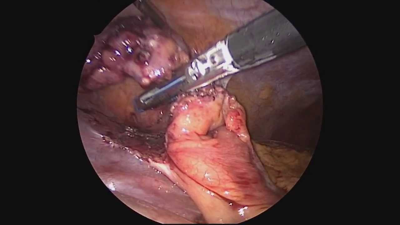
Dr. Celia Divino, Chief, Division of General Surgery at The Mount Sinai Hospital, performs a laparoscopic appendectomy. Visit the Division of General Surgery at http://bit.ly/18z944M. Click here to learn more about Dr. Celia Divino http://bit.ly/12RF0ee
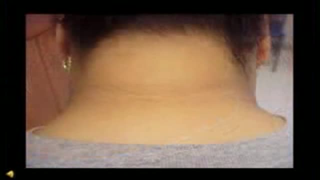
Acanthosis Nigricans Insulin Resistance
Enchondroma (Cartilage) benign tumor of the finger.

Is that knee pain just a sprain or a more serious ACL injury? Orthopedic surgeon Paul Fadale, M.D., offers tips on how to tell the difference. http://www.orthopedicsri.org/

Prosthetic hand that can feel
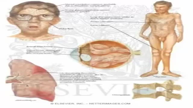
Homocystinuria is an inherited disorder that affects the metabolism of the amino acid methionine. Amino acids are the building blocks of life. Causes Homocystinuria is inherited in families as an autosomal recessive trait. This means that the child must inherit a non-working copy of the gene from each parent to be seriously affected. Homocystinuria has several features in common with Marfan syndrome, including joint and eye changes. Symptoms Newborn infants appear healthy. Early symptoms, if present, are not obvious. Symptoms may occur as mildly delayed development or failure to thrive. Increasing visual problems may lead to diagnosis of this condition. Other symptoms include: Chest deformities (pectus carinatum, pectus excavatum) Flush across the cheeks High arches of the feet Intellectual disability Knock knees Long limbs Mental disorders Nearsightedness Spidery fingers (arachnodactyly) Tall, thin build
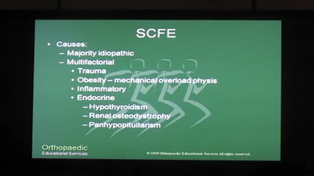
There is a strong association with obesity. In children younger than 10 years, it is associated with metabolic endocrine disorders {hypothyroidism, panhypopituitarism, hypogonadism, renal osteodystrophy, growth hormone abnormalities). SCFE is considered chronic if it has been present more than 3 weeks and acute if it has been present for 3 weeks or less. It is called "stable" if the patient can bear weight and "unstable" if the patient cannot ambulate. Unstable SCFE is associated with more complications, including avascular necrosis of the femoral head (AVN). SCFE is diagnosed by x-ray of the pelvis and bilateral hips. The underlying cause is a widened epiphyseal growth plate, due to abnormal cartilage maturation and endochondral ossification. The treatment is surgical, requiring immediate internal fixation with a single screw. Delay in treatment {> 24 hours) leads to increased AVN, SCFE progression from stable to unstable, and high risk of future degenerative arthritis. Prophylactic contralateral fixation of the unaffected hip is not routinely done in the U.S., except in patients with endocrine abnormalities.
