- Physical Examination
- Surgical Examination
- Ophthalmology
- Clinical Skills
- Orthopedics
- Surgery Videos
- Laparoscopy
- Pediatrics
- Funny Videos
- Cardiothoracic Surgery
- Nursing Videos
- Plastic Surgery
- Otorhinolaryngology
- Histology and Histopathology
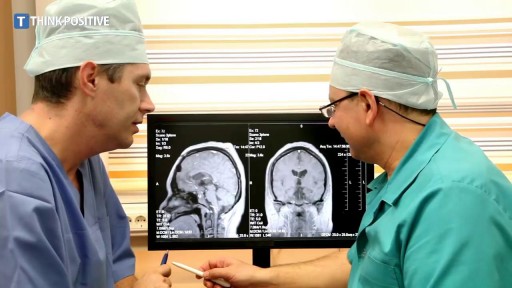
- Neurosurgery
- Dermatology
- Pediatric Surgery
- Urology
- Dentistry
- Oncology and Cancers
- Anatomy Videos
- Health and Fitness
- Radiology
- Anaesthesia
- Physical Therapy
- Pharmacology
- Interventional Radiology
- Cardiology
- Endocrinology
- Gynecology
- Emergency Medicine
- Psychiatry and Psychology
- Childbirth Videos
- General Medical Videos
- Nephrology
- Physiology
- Diet and Food Health
- Diabetes Mellitus
- Neurology
- Women Health
- Osteoporosis
- Gastroenterology
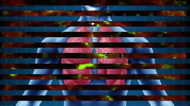
- Pulmonology
- Hematology
- Rheumatology
- Toxicology
- Nuclear Medicine
- Infectious Diseases
- Vascular Disease
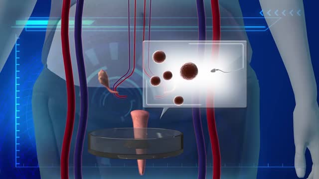
- Reproductive Health
- Burns and Wound Healing
- Other
Top videos
According to a Danish study , frequent sex may help prevent pre-eclampsia. Researchers believe it's because of a protein found in sperm that can regulate the body's immune system. Yet because the cause of preeclampsia is unknown, it's important to keep your prenatal visits and talk to your doctor about your risk.
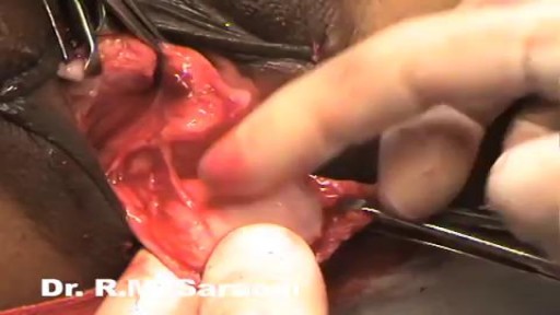
Demonstration of subcuticular or intradermal suturing technique for wound closure in the operating room.
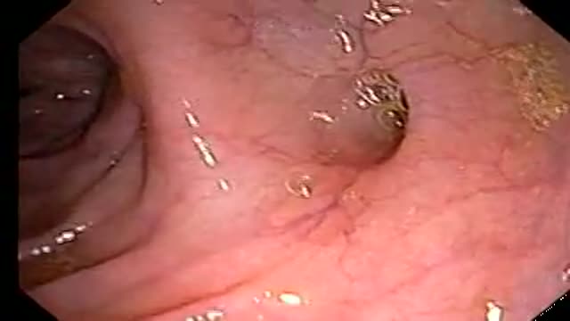
Small colon polyp (redish bump)and many diverticuli (small outpouches in wall of the colon)
Pregnancy Tips : How Early Can You Take a Blood Test for Pregnancy?
Ellie was born with a rare condition which stopped her jawbones from growing properly. At first, her parents didn't realize there was a problem, apart from the fact that her teeth were not aligned. But when she went to have braces fitted to straighten her teeth when she was 14, orthodontist Joy Hickman realized her jaw had not grown since she was eight. Over the next six years Hickman worked with a maxillofacial surgeon to transform Ellie's looks. Ellie, who is now 20, said the surgery was painful but paid almost immediate dividends. "About six months after it was my year 11 prom and it looked good." Ellie told the Daily Post the change in her appearance has been matched by an increase in confidence.
U.S. uterus transplants: experimental surgery could help infertile women get pregnant

Dr. Erica Hodgman discusses pediatric surgery at the Johns Hopkins Children's Center Pediatric General Surgery program, what common surgeries the program specializes in, what makes the program unique and her work as a pediatric surgeon. #PediatricSurgery #JohnsHopkinsChildrenCenter
Questions Answered:
0:03 Describe the pediatric general surgery division at Johns Hopkins Children's Center.
1:00 What makes this program unique?
1:31 What are some common pediatric surgery cases?
2:23 Explain your work as a pediatric general surgeon?

The Nurse Job is very hard
Cervical cancer occurs when abnormal cells on the cervix camera.gif grow out of control. The cervix is the lower part of the uterus that opens into the vagina. Cervical cancer can often be successfully treated when it's found early. It is usually found at a very early stage through a Pap test.
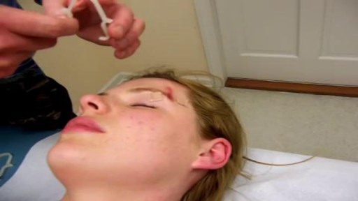
Demonstration of Burke-Baier wound closure forceps on simulated wound near eyebrow.
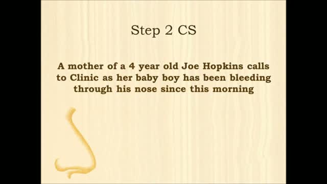
USMLE Step 2 CS - NOSE BLEEDS This is just preview video. To get full access please visit our website : www.usmletutoring.com
Rhabdomyolysis is a condition in which damaged skeletal muscle (Ancient Greek: rhabdomyo-) tissue breaks down rapidly (Greek –lysis). This damage may be caused by physical (e.g. crush injury), chemical, or biological factors. Breakdown products of damaged muscle cells are released into the bloodstream; some of these, such as the protein myoglobin, are harmful to the kidney and may lead to kidney dysfunction. The severity of the symptoms (which may include muscle pains, vomiting and confusion) depends on the extent of the muscle damage, and whether kidney failure develops. The mainstay of treatment is generous intravenous fluids, but could include dialysis or hemofiltration.
Rhabdomyolysis and its complications are significant problems for those injured in disasters such as earthquakes and bombing. Relief efforts in areas struck by earthquakes often include medical teams with skills and equipment for treatment of survivors with rhabdomyolysis. The disease and its mechanisms were first fully elucidated during the Blitz of London in 1941.
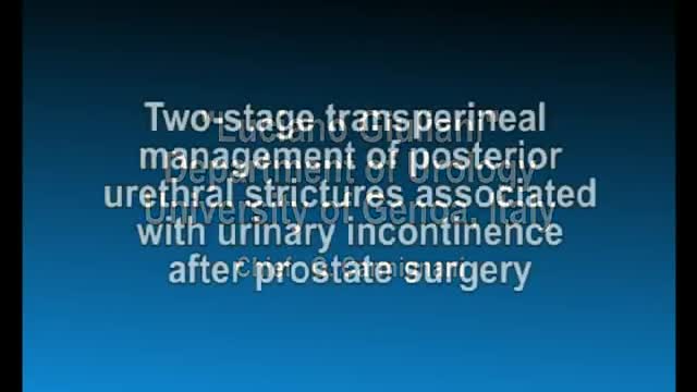
Posterior Urethral Strictures Associated with Urinary Incontinence after Prostatectomy Management
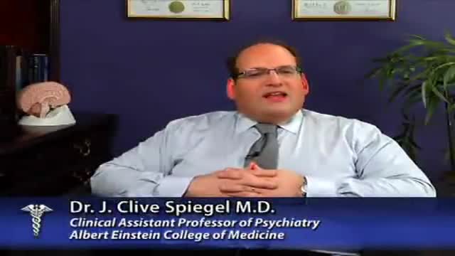
Antisocial personality disorder (ASPD) is defined by the American Psychiatric Association's Axis II (personality disorders) of the Diagnostic and Statistical Manual (DSM-IV-TR) as "a pervasive pattern of disregard for, and violation of, the rights of others that begins in childhood or early adolescence and continues into adulthood." Antisocial personality disorder is sometimes wrongly referred to as psychopathy or sociopathy. Currently, neither psychopathy nor sociopathy are valid diagnoses described in the Diagnostic and Statistical Manual of Mental Disorders, and the ICD-10 of the World Health Organization also lacks psychopathy as a diagnostic disorder. Psychopathy is normally seen as a subset of the antisocial personality disorder, but Blair believes that the antisocial personality disorder and psychopathy may be separate conditions altogether.
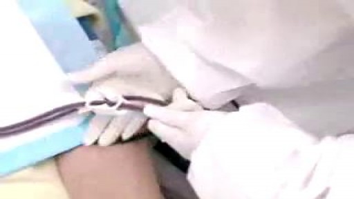
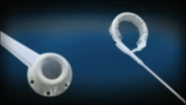
Preventing Hemodialysis Catheters Problems
A good starting point for any scientist in any field is to recognize that there is much we do not know. We do not know, for example, why there is more matter than antimatter in the universe. We do not know very well how the evolution of the dinosaurs filtered out. And, perhaps most surprising of all is that we do not know very well how many organs the human body has or what all its functions are.
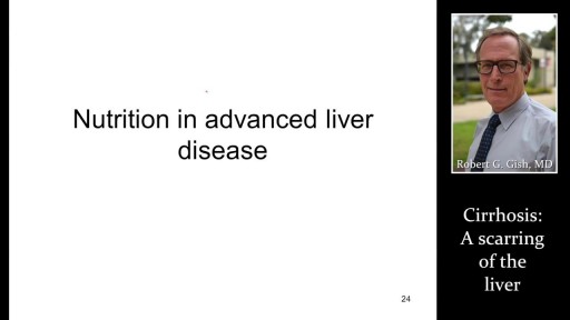
Hepatitis and chronic alcohol abuse are frequent causes. Liver damage caused by cirrhosis can't be undone, but further damage can be limited. Initially patients may experience fatigue, weakness, and weight loss. During later stages, patients may develop jaundice (yellowing of the skin), gastrointestinal bleeding, abdominal swelling, and confusion. Treatments focus on the underlying cause. In advanced cases, a liver transplant may be needed.
Handal Plastic Surgery at the Sanctuary Surgery Center is the leading cosmetic surgery center of the Southeast Florida region, providing excellent consultation, surgery, and post operative services. Headed by Doctor Arthur G. Handal, top plastic & cosmetic surgeon in Boca Raton, the professional staff of the Sanctuary Surgery Center offers the best in patient care.
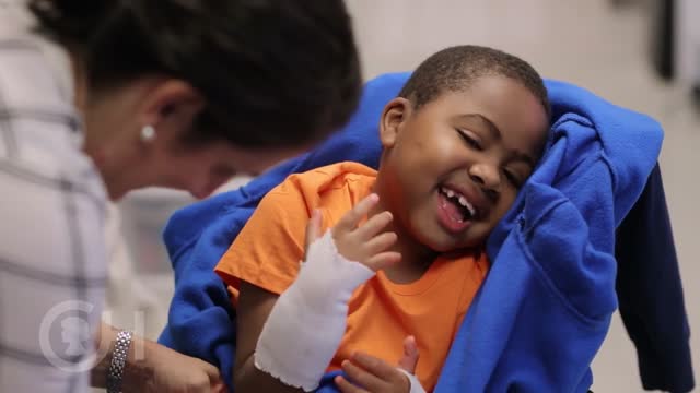
Surgeons at The Children’s Hospital of Philadelphia were the first to perform a bilateral hand transplant on a child. Our research and work in this groundbreaking field of medicine led us to establish the Hand Transplantation Program. Combining the expertise of the Penn Transplant Institute and the Hospital’s Division of Plastic and Reconstructive Surgery and Division of Orthopedics, the program aims to improve quality of life for children who may benefit from this procedure. This is Zion, one year after the surgery
