- Physical Examination
- Surgical Examination
- Ophthalmology
- Clinical Skills
- Orthopedics
- Surgery Videos
- Laparoscopy
- Pediatrics
- Funny Videos
- Cardiothoracic Surgery
- Nursing Videos
- Plastic Surgery
- Otorhinolaryngology
- Histology and Histopathology
- Neurosurgery
- Dermatology
- Pediatric Surgery
- Urology
- Dentistry
- Oncology and Cancers
- Anatomy Videos
- Health and Fitness
- Radiology
- Anaesthesia
- Physical Therapy
- Pharmacology
- Interventional Radiology
- Cardiology
- Endocrinology
- Gynecology
- Emergency Medicine
- Psychiatry and Psychology
- Childbirth Videos
- General Medical Videos
- Nephrology
- Physiology
- Diet and Food Health
- Diabetes Mellitus
- Neurology
- Women Health
- Osteoporosis
- Gastroenterology
- Pulmonology
- Hematology
- Rheumatology
- Toxicology
- Nuclear Medicine
- Infectious Diseases
- Vascular Disease
- Reproductive Health
- Burns and Wound Healing
- Other
Top videos
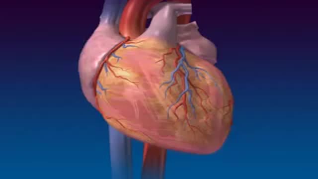
Sectioned Heart
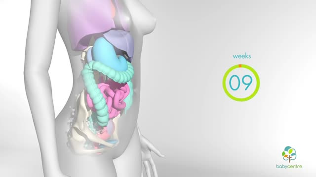
In your first few months of pregnancy, hormones flood your body. Your baby is still tiny but already your body is changing. Your breasts start to swell and may feel tender. Tiredness, nausea and frequent trips to the loo are common pregnancy symptoms.

Watch that video to know How to Get Pregnant Fast and Easy
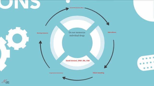
Remembering Medications & The Body Systems Affected
A Cesarean section (C-section) is surgery to deliver a baby. The baby is taken out through the mother's abdomen. In the United States, almost one in three women has their babies this way. Some C-sections are planned, but many are done when unexpected problems happen during delivery. Reasons for a C-section may include
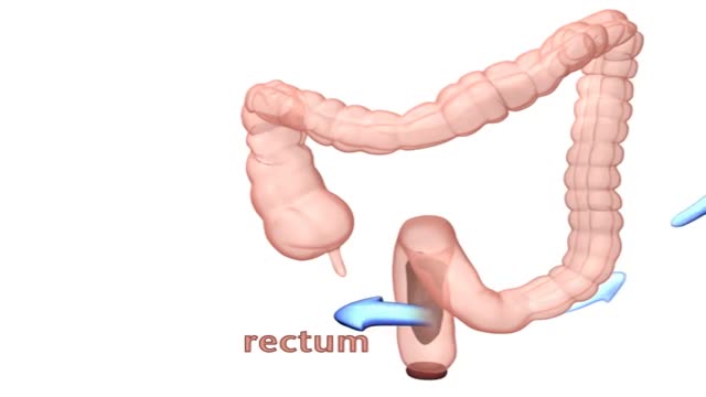
Kegel exercises strengthen the pelvic floor muscles, which support the uterus, bladder, small intestine and rectum. You can do Kegel exercises, also known as pelvic floor muscle training, just about anytime. Start by understanding what Kegel exercises can do for you — then follow step-by-step instructions for contracting and relaxing your pelvic floor muscles.

http://www.landging.com/breast-enlargement-animation.html
Breast enlargement product animation designed for TV shopping.

http://www.landging.com/expo2010_case_2.html
Airplane perspective animation, 3d aviation animation, developed for Expo 2010 Shanghai Aviation Pavilion.
Encopresis is a problem that children age four or older can develop due to chronic (long-term) constipation. With constipation, children have fewer bowel movements than normal, and the bowel movements they do have can be hard, dry, and difficult to pass. The child may avoid using the bathroom to avoid discomfort.

Duke Sports Medicine Specialists Jocelyn Wittstein, MD, Janna Fonseca, ATC, and Michael Messer ,PT, present on Soccer Injury Prevention including Concussion Management and the 11+ program that significantly reduces ACL tear rates in soccer.

This video shows the heart transplant surgery

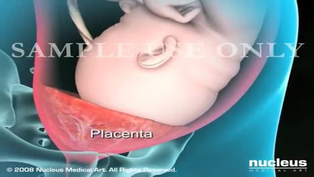
Perineal repair after episiotomy or spontaneous obstetric laceration is one of the most common surgical procedures. Potential sequelae of obstetric perineal lacerations include chronic perineal pain,1 dyspareunia,2 and urinary and fecal incontinence.3–5 Few studies of laceration repair techniques exist to support the development of an evidence-based approach to perineal repair. This article discusses a repair method that emphasizes anatomic detail, with the expectation that an anatomically correct perineal repair may result in a better long-term functional outcome.
Here are seven ways to start reining in your risks today, before a stroke has the chance to strike. Lower blood pressure. ... Lose weight. ... Exercise more. ... Drink — in moderation. ... Treat atrial fibrillation. ... Treat diabetes. ... Quit smoking.

Cosmetic facial plastic surgery is surgery performed to enhance visual appearance of the facial structures and features. Common procedures include facelifts, eye lifts, rhinoplasty, chin and cheek implants, liposuction, and procedures to correct facial wrinkles.

Handal Plastic Surgery at the Sanctuary Surgery Center is the leading cosmetic surgery center of the Southeast Florida region, providing excellent consultation, surgery, and post operative services. Headed by Doctor Arthur G. Handal, top plastic & cosmetic surgeon in Boca Raton, the professional staff of the Sanctuary Surgery Center offers the best in patient care.

If you have a blocked artery, your doctor may need to open the blockage and restore blood flow using a small mesh tube called a stent. The stent is inserted in your artery during an angioplasty procedure. Until now, stents were permanent. Now there is a fully dissolving stent available to treat blockages.

Surgeons at The Children’s Hospital of Philadelphia were the first to perform a bilateral hand transplant on a child. Our research and work in this groundbreaking field of medicine led us to establish the Hand Transplantation Program. Combining the expertise of the Penn Transplant Institute and the Hospital’s Division of Plastic and Reconstructive Surgery and Division of Orthopedics, the program aims to improve quality of life for children who may benefit from this procedure. This is Zion, one year after the surgery
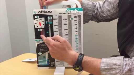
If you’re like me, you probably hook your chest tube up to a Pleur-Evac, put it on the ground, then back away slowly. Who knows what goes on in that mysterious bubbling white box? Hopefully this will post shed some light. Isn’t this just a container for stuff that comes out of the chest? Why does it look so complicated? It’s complicated because the detection/collection of air and fluid require different setups. Most commercial models also allow you to hook the drainage system to wall suction, so you can quickly evacuate the pleural space. This requires its own setup. Because of the need to juggle air, fluid and suction, the most common commercial system includes 3 distinct chambers. If you were to simplify the device, or build one out of spare bottles and tubes, it might look like this:

What Causes Ulcers? No single cause has been found for ulcers. However, it is now clear that an ulcer is the end result of an imbalance between digestive fluids in the stomach and duodenum. Most ulcers are caused by an infection with a type of bacteria called Helicobacter pylori (H. pylori). Factors that can increase your risk for ulcers include: Use of painkillers called nonsteroidal anti-inflammatory drugs (NSAIDs), such as aspirin, naproxen (Aleve, Anaprox, Naprosyn, and others), ibuprofen (Motrin, Advil, some types of Midol, and others), and many others available by prescription; even safety-coated aspirin and aspirin in powered form can frequently cause ulcers. Excess acid production from gastrinomas, tumors of the acid producing cells of the stomach that increases acid output (seen in Zollinger-Ellison syndrome) Excessive drinking of alcohol Smoking or chewing tobacco Serious illness Radiation treatment to the area What Are the Symptoms of an Ulcer? An ulcer may or may not have symptoms. When symptoms occur, they may include: A gnawing or burning pain in the middle or upper stomach between meals or at night Bloating Heartburn Nausea or vomiting In severe cases, symptoms can include: Dark or black stool (due to bleeding) Vomiting blood (that can look like "coffee-grounds") Weight loss Severe pain in the mid to upper abdomen
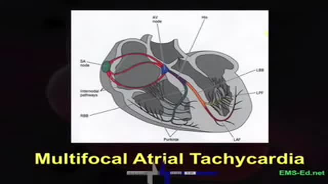
EKG Interpretation Part 3
