- Physical Examination
- Surgical Examination
- Ophthalmology
- Clinical Skills
- Orthopedics
- Surgery Videos
- Laparoscopy
- Pediatrics
- Funny Videos
- Cardiothoracic Surgery
- Nursing Videos
- Plastic Surgery
- Otorhinolaryngology
- Histology and Histopathology
- Neurosurgery
- Dermatology
- Pediatric Surgery
- Urology
- Dentistry
- Oncology and Cancers
- Anatomy Videos
- Health and Fitness
- Radiology
- Anaesthesia
- Physical Therapy
- Pharmacology
- Interventional Radiology
- Cardiology
- Endocrinology
- Gynecology
- Emergency Medicine
- Psychiatry and Psychology
- Childbirth Videos
- General Medical Videos
- Nephrology
- Physiology
- Diet and Food Health
- Diabetes Mellitus
- Neurology
- Women Health
- Osteoporosis
- Gastroenterology
- Pulmonology
- Hematology
- Rheumatology
- Toxicology
- Nuclear Medicine
- Infectious Diseases
- Vascular Disease
- Reproductive Health
- Burns and Wound Healing
- Other
Top videos
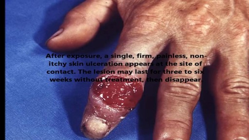
STDs are infections that are transmitted during vaginal, anal, and oral sex. They are very common and many people who have them don't show any symptoms.

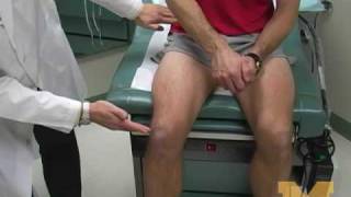
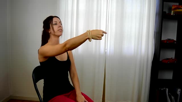
Dr. Yeong Kwok speaks about knee pain and demonstrates a stretch designed to treat tendonitis.
The cause for TS is unknown. Early research suggested that TS is an inherited condition (often, the person's near or distant relatives have had some form of transient or chronic tic disorder or associated symptoms). Recent studies point to a combination of environmental and genetic factors as a cause of the disorder. The specific genes involved in the development of TS are still being investigated. Studies suggest that TS has a neurological basis and results from an abnormality which affects the brain's metabolism of certain neurotransmitters (chemicals in the brain that regulate behavior.) Current research being funded by the Tourette Syndrome Association (TSA) will help provide more information about the causes and genetic factors of TS.
There can be a number of psychological triggers that cause nightmares in adults. For example, anxiety and depression can cause adult nightmares. Post-traumatic stress disorder (PTSD) also commonly causes people to experience chronic, recurrent nightmares. Nightmares in adults can be caused by certain sleep disorders

Polyarteritis nodosa Email this page to a friend Email this page to a friend Facebook Twitter Google+ Polyarteritis nodosa is a serious blood vessel disease. The small and medium-sized arteries become swollen and damaged. Causes Arteries are the blood vessels that carry oxygen-rich blood to organs and tissues. The cause of polyarteritis nodosa is unknown. The condition occurs when certain immune cells attack the affected arteries. More adults than children get this disease. The tissues that are fed by the affected arteries do not get the oxygen and nourishment they need. Damage occurs as a result. People with active hepatitis B or hepatitis C may develop this disease.

Proximal Hypospadias repaired by Tube Onaly Urethroplasty

How to open a glass ampoule

Demonstration of simple interrupted suturing technique for laceration repair.
This system treats type 2 diabetes by promoting weight loss.
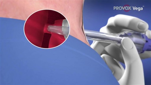
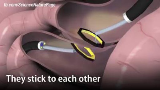
A voice prosthesis (plural prostheses) is an artificial device, usually made of silicone that is used to help laryngectomized patients to speak. During a total laryngectomy, the entire voice box (larynx) is removed and the windpipe (trachea) and food pipe (esophagus) are separated from each other.

Cognitive impairment is seen in over half of people with multiple sclerosis. In this video I review factors that can contribute to multiple sclerosis brain fog, ways to assess cognition, and tips to improve thinking and memory.

Mommy Makeover plastic surgery in NYC and is a fairly new phenomena. This video, from 5thavenue surgery; http://www.5thavesurgery.com, goes through a case study of a patient getting plastic surgery in NYC. Check out what a Mommy Makeover can do for your body and what Plastic Surgery can do for you.

There are four major blood groups determined by the presence or absence of two antigens – A and B – on the surface of red blood cells: Group A – has only the A antigen on red cells (and B antibody in the plasma) Group B – has only the B antigen on red cells (and A antibody in the plasma) Group AB – has both A and B antigens on red cells (but neither A nor B antibody in the plasma) Group O – has neither A nor B antigens on red cells (but both A and B antibody are in the plasma)
Multiple sclerosis (MS) is a potentially disabling disease of the brain and spinal cord (central nervous system). In MS, the immune system attacks the protective sheath (myelin) that covers nerve fibers and causes communication problems between your brain and the rest of your body. Eventually, the disease can cause the nerves themselves to deteriorate or become permanently damaged. Signs and symptoms of MS vary widely and depend on the amount of nerve damage and which nerves are affected. Some people with severe MS may lose the ability to walk independently or at all, while others may experience long periods of remission without any new symptoms. There's no cure for multiple sclerosis. However, treatments can help speed recovery from attacks, modify the course of the disease and manage symptoms
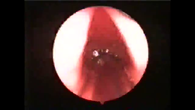
This clip shows an endoscopic view of the vocal cords with the endotracheal tube in place. The patient was intubated by Dr. Khaled Soliman and photographed by Dr. Mohammed Wahba.

HD Gynecomastia Surgery
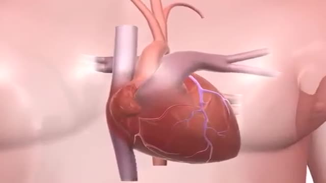
Uncontrolled high blood pressure can lead to stroke by damaging and weakening your brain's blood vessels, causing them to narrow, rupture or leak. High blood pressure can also cause blood clots to form in the arteries leading to your brain, blocking blood flow and potentially causing a stroke. Dementia.
ADH's job is to act on the kidneys to promote water reabsorption. In this lesson, we'll compare and contrast diabetes insipidus, or DI, in which there is too little ADH, and syndrome of inappropriate antidiuretic hormone secretion , or SIADH, in which there is too much ADH.
Vertigo is a sensation of spinning. If you have these dizzy spells, you might feel like you are spinning or that the world around you is spinning. Causes of Vertigo Vertigo is often caused by an inner ear problem. Some of the most common causes include: BPPV. These initials stand for benign paroxysmal positional vertigo. BPPV occurs when tiny calcium particles (canaliths) clump up in canals of the inner ear. The inner ear sends signals to the brain about head and body movements relative to gravity. It helps you keep your balance. BPPV can occur for no known reason and may be associated with age. Meniere's disease. This is an inner ear disorder thought to be caused by a buildup of fluid and changing pressure in the ear. It can cause episodes of vertigo along with ringing in the ears (tinnitus) and hearing loss. Vestibular neuritis or labyrinthitis. This is an inner ear problem usually related to infection (usually viral). The infection causes inflammation in the inner ear around nerves that are important for helping the body sense balance
