- Physical Examination
- Surgical Examination
- Ophthalmology
- Clinical Skills
- Orthopedics
- Surgery Videos
- Laparoscopy
- Pediatrics
- Funny Videos
- Cardiothoracic Surgery
- Nursing Videos
- Plastic Surgery
- Otorhinolaryngology
- Histology and Histopathology
- Neurosurgery
- Dermatology
- Pediatric Surgery
- Urology
- Dentistry
- Oncology and Cancers
- Anatomy Videos
- Health and Fitness
- Radiology
- Anaesthesia
- Physical Therapy
- Pharmacology
- Interventional Radiology
- Cardiology
- Endocrinology
- Gynecology
- Emergency Medicine
- Psychiatry and Psychology
- Childbirth Videos
- General Medical Videos
- Nephrology
- Physiology
- Diet and Food Health
- Diabetes Mellitus
- Neurology
- Women Health
- Osteoporosis
- Gastroenterology
- Pulmonology
- Hematology
- Rheumatology
- Toxicology
- Nuclear Medicine
- Infectious Diseases
- Vascular Disease
- Reproductive Health
- Burns and Wound Healing
- Other
Top videos
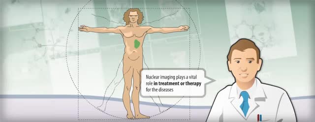
Nuclear medicine is a branch of medical imaging that uses small amounts of radioactive material to diagnose and determine the severity of or treat a variety of diseases, including many types of cancers, heart disease, gastrointestinal, endocrine, neurological disorders and other abnormalities within the body.
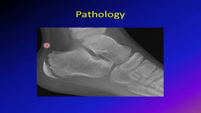
Sever's disease (also known as calcaneal apophysitis) is a type of bone injury in which the growth plate in the lower back of the heel, where the Achilles tendon (the heel cord that attaches to the growth plate) attaches, becomes inflamed and causes pain.


http://plantar-fasciitis-solution.info-pro.co Pain In Arch Of Foot, Severe Heel Pain, Best Running Shoes For Plantar Fasciitis, Foot Pain Heel Know the Symptoms of Plantar Fasciitis An injury to the plantar fascia can manifest in different ways. Initially, it may be a gradual pain that can progressively become worse, especially if the injured foot remains in active use. Sometimes, the pain from plantar fasciitis can be quite severe and seem like the stab of a knife of a sharp and sudden cut. The pain of plantar fasciitis may also occur more frequently after injured feet have been at rest for a while. For instance, after a person wakes up and tries to use his or her feet, pain may be experienced. It can be dangerous to ignore pain that is associated with the feet or any pain felt in the body. Sometimes, symptoms of plantar fasciitis include more subtle pain that may appear as a throbbing sensation which may be radial in nature or isolated to a particular part of the foot. If the pain from plantar fasciitis starts off mildly and is ignored, continued use of the affected foot or feet will cause further damage. The pain from plantar fasciitis is debilitating and it is essential that treatment is sought immediately. get instant plantar fasciitis pain relief in just 5 minutes! click here. http://plantar-fasciitis-solution.info-pro.co
A giant abdominal wall hernia can develop from an existing ventral or incisional hernia, sometimes arising after one or more failed repair attempts. These hernias may also result from a traumatic injury where the abdomen was required to be left open and healing was delayed. In giant abdominal wall hernias, multiple loops of intestines and sometimes other abdominal organs reside within the hernia sac. The abdominal wall muscles then become conditioned to this and retract reducing the available space inside the abdomen.
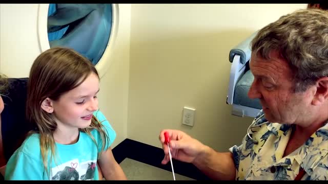
Strep throat is a bacterial infection that can make your throat feel sore and scratchy. Strep throat accounts for only a small portion of sore throats. If untreated, strep throat can cause complications, such as kidney inflammation or rheumatic fever. Rheumatic fever can lead to painful and inflamed joints, a specific type of rash or heart valve damage. Strep throat is most common in children, but it affects people of all ages. If you or your child has signs or symptoms of strep throat, see your doctor for prompt testing and treatment.
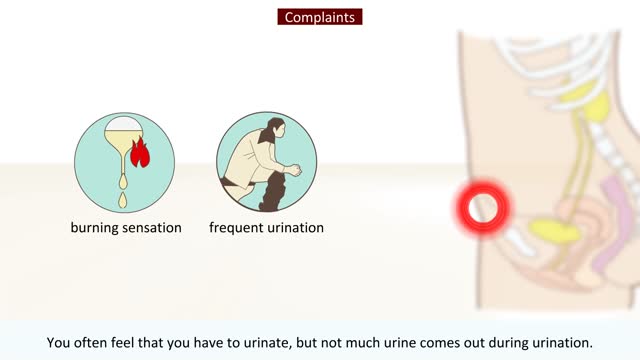
Most women are put on a 3 to 5 day antibiotic. Men might be put on an antibiotic for 7 to 14 days. While symptoms usually clear up around three days after antibiotic treatment, it can take up to five days for all the bacteria in your urinary tract to die off. It may take even longer for men.
Traditionally, the appendix is removed through an incision in the right lower abdominal wall. In most laparoscopic appendectomies, surgeons operate through 3 small incisions (each ¼ to ½ inch) while watching an enlarged image of the patient's internal organs on a television monitor.

The CSICU rounds are an opportunity for residents to come together with attendings and review all the patients in the ICU.
Cedars-Sinai is committed to educating exceptional cardiothoracic surgeons through outstanding personal mentorship, operative training and research leadership. Residents of the Thoracic Surgery—Integrated Residency at Cedars-Sinai will be part of an incredibly rich, academic environment—each year our research and thought leadership features in hundreds of publications in journals including Nature, New England Journal of Medicine, JAMA, Lancet and leading specialty journals.
Learn more about the Cedars-Sinai Thoracic Surgery—Integrated Residency: https://ceda.rs/3UDrZFL
Connect with us:
https://twitter.com/CedarsSinai
https://www.facebook.com/CedarsSinai
https://www.instagram.com/CedarsSinai
Cedars-Sinai is a leader in providing high-quality healthcare encompassing primary care, specialized medicine and research. Since 1902, Cedars-Sinai has evolved to meet the needs of one of the most diverse regions in the nation, setting standards in quality and innovative patient care, research, teaching and community service. Today, Cedars- Sinai is known for its national leadership in transforming healthcare for the benefit of patients. Cedars-Sinai impacts the future of healthcare by developing new approaches to treatment and educating tomorrow’s health professionals. Additionally, Cedars-Sinai demonstrates a commitment to the community through programs that improve the health of its most vulnerable residents.

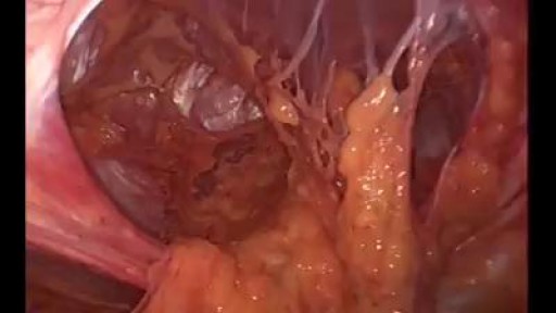
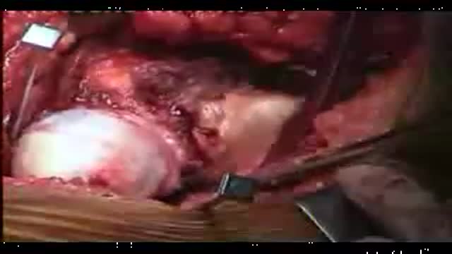
Hydatid cysts in retroperitoneal region in transit to the thorax
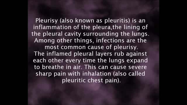
Pleurisy is a condition in which the pleura — a membrane consisting of a layer of tissue that lines the inner side of the chest cavity and a layer of tissue that surrounds the lungs — becomes inflamed. Also called pleuritis, pleurisy causes sharp chest pain (pleuritic pain) that worsens during breathing. A variety of underlying conditions can cause pleurisy. Treatment of pleurisy involves pain control and treating the underlying condition.
Dr. Vijay Bose is awarded by Sri. P. Chidambaram Honorable Minister of Finance for Young Achiver Winning Award Progarm.
While the incidence of most sports-related injuries has been holding steady for the past two decades, injuries to the anterior cruciate ligament (ACL) continue to increase significantly, particularly in female athletes. In fact, on many college teams, as many as 30 to 50 percent of young women have had an ACL injury during their high school careers in certain sports, such as basketball, soccer and gymnastics.
Watch pediatric orthopedic surgeons at Akron Children's Hospital perform arthroscopic surgery to replace a young athlete's ACL
Another video of Dr.Vijay C. Bose from Apollo Speciality Hospital chennai perform Birmingham Hip Resurfacing Surgery procedure for a case of Avascular necrosis.The NCP ( Neck Capsule Preserving) approach is being used. Total hip replacement, hip resurfacing simply shaves and caps a few centimeters of bone within the joint. The bone-conserving approach of the Birmingham Hip Resurfacing System.
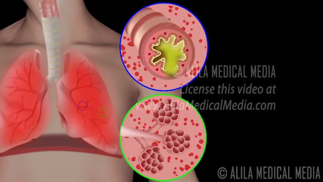
Chronic obstructive pulmonary disease Email this page to a friend Print Facebook Twitter Google+ Chronic obstructive pulmonary disease (COPD) is a common lung disease. Having COPD makes it hard to breathe. There are two main forms of COPD: Chronic bronchitis, which involves a long-term cough with mucus Emphysema, which involves damage to the lungs over time Most people with COPD have a combination of both conditions. Causes Smoking is the main cause of COPD. The more a person smokes, the more likely that person will develop COPD. But some people smoke for years and never get COPD. In rare cases, nonsmokers who lack a protein called alpha-1 antitrypsin can develop emphysema. Emphysema Other risk factors for COPD are: Exposure to certain gases or fumes in the workplace Exposure to heavy amounts of secondhand smoke and pollution Frequent use of a cooking fire without proper ventilation Symptoms Symptoms may include any of the following: Cough, with or without mucous Fatigue Many respiratory infections Shortness of breath (dyspnea) that gets worse with mild activity Trouble catching one's breath Wheezing Because the symptoms develop slowly, some people may not know that they have COPD.
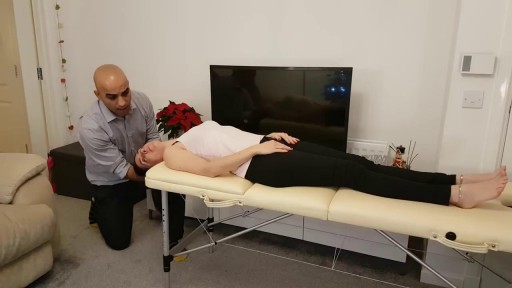
The Epley maneuver is a series of movements, normally carried out on a person by a doctor, to relieve the symptoms of BPPV. Research has found it to be an easy, safe, and effective treatment for the condition in both the long- and short-term. The Epley maneuver is sometimes called the particle repositioning maneuver or the canalith repositioning maneuver. These names are used because the maneuver involves a series of movements that help to reposition crystals in a person's ear that may cause feelings of dizziness. Repositioning the crystals helps to relieve the person's dizziness and nausea.
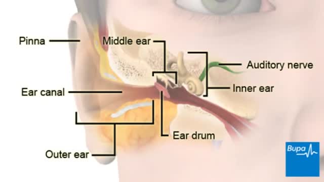
Labyrinthitis is a mild, often self-limited condition characterized by vertigo, tinnitus, nausea, and a loss of balance. The disorder often follows a viral illness (eg, influenza). Labyrinthitis may also be caused by trauma, bacterial infection, allergies, benign tumors, and certain medications .
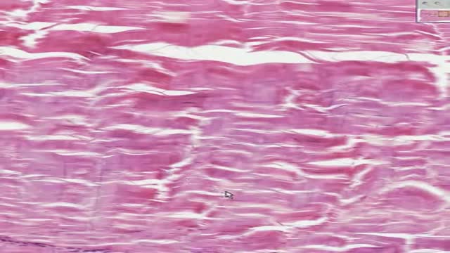
Histology of Dense Regular Connective Tissue
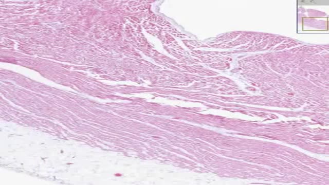
Histology of Heart Cardiac Muscle
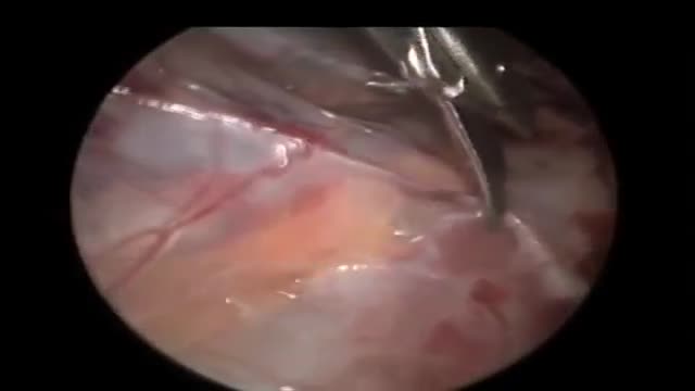
Laparoscopy seems to offer a safe and reliable diagnostic and therapeutic option to patients with impalpable testes. Intra-abdominal dissection allows more testes to be brought down to the scrotum. The procedure is best viewed as laparoscopy-assisted, as Orchidopexy has to be done in a conventional manner.
