- Physical Examination
- Surgical Examination
- Ophthalmology
- Clinical Skills
- Orthopedics
- Surgery Videos
- Laparoscopy
- Pediatrics
- Funny Videos
- Cardiothoracic Surgery
- Nursing Videos
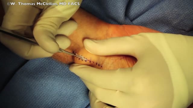
- Plastic Surgery
- Otorhinolaryngology
- Histology and Histopathology
- Neurosurgery
- Dermatology
- Pediatric Surgery
- Urology
- Dentistry
- Oncology and Cancers
- Anatomy Videos
- Health and Fitness
- Radiology
- Anaesthesia
- Physical Therapy
- Pharmacology
- Interventional Radiology
- Cardiology
- Endocrinology
- Gynecology
- Emergency Medicine
- Psychiatry and Psychology
- Childbirth Videos
- General Medical Videos
- Nephrology
- Physiology
- Diet and Food Health
- Diabetes Mellitus
- Neurology
- Women Health
- Osteoporosis
- Gastroenterology
- Pulmonology
- Hematology
- Rheumatology
- Toxicology
- Nuclear Medicine
- Infectious Diseases
- Vascular Disease
- Reproductive Health
- Burns and Wound Healing
- Other
Top videos
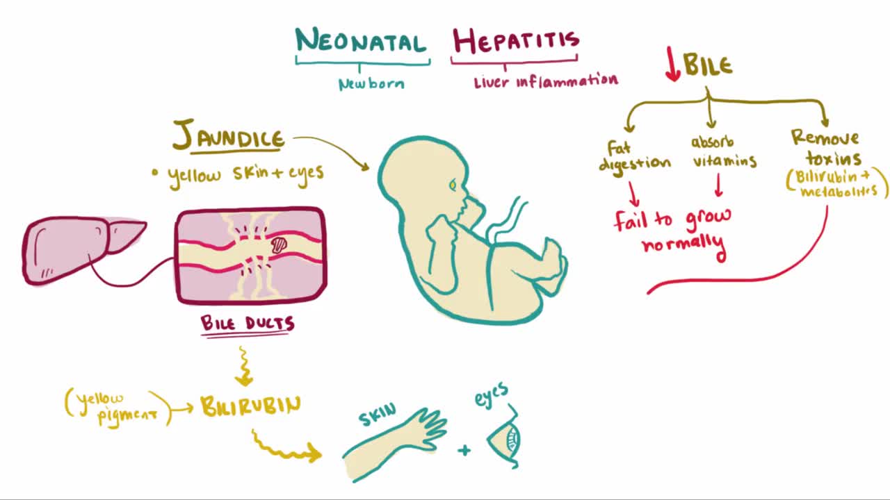
What is neonatal hepatitis? Neonatal hepatitis is an inflammation of an infant's liver just after birth, sometimes this inflammation is due to a virus but in most cases the cause is unknown, or idiopathic
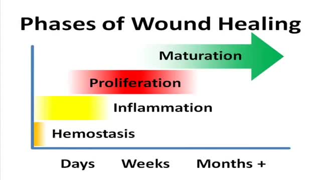
A video showing the phases of normal wound healing
Osteoporosis is a disease in which bones become brittle and fragile due to low bone mass and bone tissue loss. It's the most common type of bone disease, according to the National Institutes of Health (NIH), and increases your risk of fractures, particularly of the hips, spine, and wrists. Prevalence In the United States, nearly 54 million people ages 50 and older were living with osteoporosis or osteopenia (low bone mass ) in 2010, according to a 2014 article in the Journal of Bone and Mineral Research. More specifically, 10.2 million adults had osteoporosis, and 43.4 million adults had osteopenia, which puts a person at high risk for osteoporosis.
USMLE Step 2 CS - Pain Seeking This is just preview video. To get full access please visit our website : www.usmletutoring.com
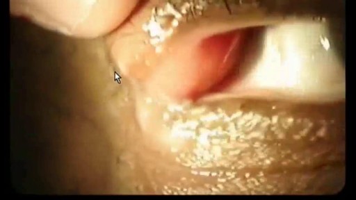
A stye (also called a hordeolum) is a small, red, painful lump that grows from the base of your eyelash or under the eyelid. Most styes are caused by a bacterial infection. There are two kinds of styes: External hordeolum: A stye that begins at the base of your eyelash. Most are caused by an infection in the hair follicle. It might look like a pimple. Internal hordeolum: A stye inside your eyelid. Most are caused by an infection in an oil-producing gland in your eyelid.
Paracentesis is a procedure to take out fluid that has collected in the belly (peritoneal fluid). This fluid buildup is called ascites camera.gif. Ascites may be caused by infection, inflammation, an injury, or other conditions, such as cirrhosis or cancer. The fluid is taken out using a long, thin needle put through the belly. The fluid is sent to a lab and studied to find the cause of the fluid buildup. Paracentesis also may be done to take the fluid out to relieve belly pressure or pain in people with cancer or cirrhosis.
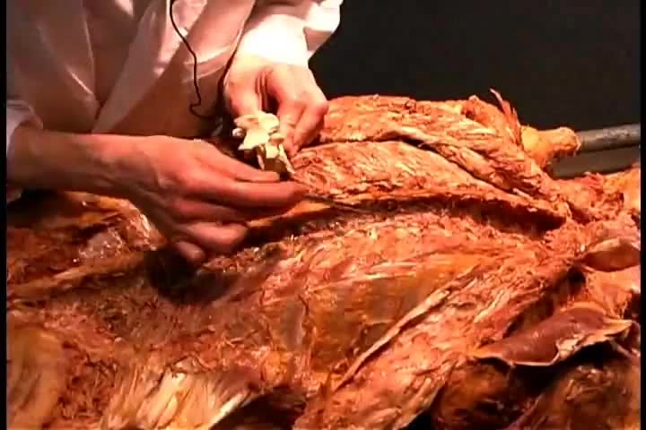
Anatomy of Back Muscles and Spinal Cord
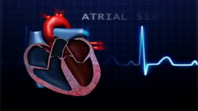
atrial septal defect (ASD) is a hole in the wall between the two upper chambers of your heart (atria). The condition is present from birth (congenital). Small atrial septal defects may close on their own during infancy or early childhood. Large and long-standing atrial septal defects can damage your heart and lungs. Small defects may never cause a problem and may be found incidentally. An adult who has had an undetected atrial septal defect for decades may have a shortened life span from heart failure or high blood pressure that affects the arteries in the lungs (pulmonary hypertension). Surgery may be necessary to repair atrial septal defects to prevent complications
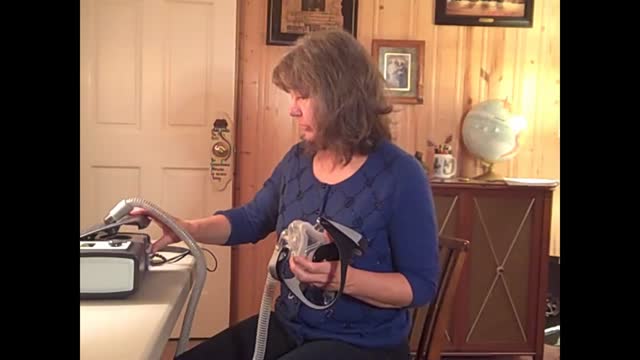
CPAP is a treatment that uses mild air pressure to keep your breathing airways open. It involves using a CPAP machine that includes a mask or other device that fits over your nose or your nose and mouth, straps to position the mask, a tube that connects the mask to the machine’s motor, and a motor that blows air into the tube. CPAP is used to treat sleep-related breathing disorders including sleep apnea. It also may be used to treat preterm infants who have underdeveloped lungs.

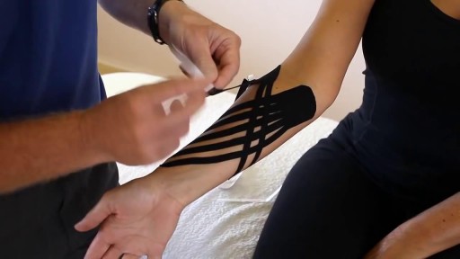
While it is unclear whether high heel shoes may or may not cause back pain, it is common for high heels to exacerbate an already present spinal condition. ... This pain in the back may also result from foot or leg fatigue that results from wearing these shoes and this can affect whole body mechanics.

DermaClinix, The world's leading hair Transplantation and aesthetic dermatology clinic that offers the best hair transplant in Chennai. The clinic is fully equipped with infrastructure and the ultra-modern devices in the field of cosmetology and hair restoration. At DermaClinx Chennai offers a number of treatments including chemical peels for acne, pigmentation treatment, glow facials, hair growth therapies and many more. Hair transplant Doctors at DermaClinix Nungambakkam, Chennai have more than 12 years of experience in this field & holds the International accreditations and member of ISHRS (USA). Visit us today to know more about the hair growth treatments. https://www.dermatologistchennai.in/hair-transplant-surgeon-in-chennai.php Address: No:19 1st Floor TMA Tower Dr.Thirumurthy Nagar main road, Nungambakkam, Chennai, Tamil Nadu 600034 Call us at - +918939636222, +91 89398 81919 For more: Website - https://www.hairtransplantchennai.org/ Email - enquiry@hairtransplantchennai.org

An orgasm is a feeling of intense sexual pleasure that happens during sexual activity. It's sometimes called "coming" or "climaxing". Both men and women have orgasms.

Microsoft HoloLens. Medical Education
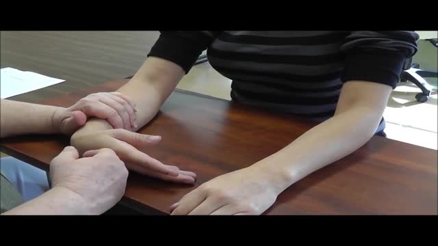
Carpal tunnel syndrome is a hand condition that causes numbness, tingling and other symptoms. Carpal tunnel syndrome is caused by a pinched nerve in your wrists A number of factors can contribute to carpal tunnel syndrome, including the anatomy of your wrist, certain underlying health problems and possibly patterns of hand use. Bound by bones and ligaments, the carpal tunnel is a narrow passageway located on the palm side of your wrist. This tunnel protects a main nerve to your hand and the nine tendons that bend your fingers. Compression of the nerve produces the numbness, tingling and, eventually, hand weakness that characterize carpal tunnel syndrome.

The exact cause of schizophrenia isn't known, but genetics, environment, and imbalanced brain chemicals may play a role. Schizophrenia is characterized by abnormal social behavior. In severe cases, patients may see or hear things that aren't real. Treatment is usually lifelong and often involves a combination of medications and psychological and social therapy.
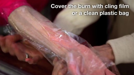
A burn is tissue damage that results from scalding, overexposure to the sun or other radiation, contact with flames, chemicals or electricity, or smoke inhalation. Is it a major or minor burn? Call 911 or seek immediate care for major burns, which: Are deep Cause the skin to be dry and leathery May appear charred or have patches of white, brown or black Are larger than 3 inches (about 8 centimeters) in diameter or cover the hands, feet, face, groin, buttocks or a major joint A minor burn that doesn't require emergency care may involve: Superficial redness similar to a sunburn Pain Blisters An area no larger than 3 inches (about 8 centimeters) in diameter Treating major burns Until emergency help arrives: Protect the burned person from further harm. If you can do so safely, make sure the person you're helping is not in contact with the source of the burn. For electrical burns, make sure the power source is off before you approach the burned person. Make certain that the person burned is breathing. If needed, begin rescue breathing if you know how. Remove jewelry, belts and other restrictive items, especially from around burned areas and the neck. Burned areas swell rapidly. Cover the area of the burn. Use a cool, moist bandage or a clean cloth. Don't immerse large severe burns in water. Doing so could cause a serious loss of body heat (hypothermia). Elevate the burned area. Raise the wound above heart level, if possible. Watch for signs of shock. Signs and symptoms include fainting, pale complexion or breathing in a notably shallow fashion. Treating minor burns For minor burns: Cool the burn. Hold the burned area under cool (not cold) running water or apply a cool, wet compress until the pain eases. Remove rings or other tight items from the burned area. Try to do this quickly and gently, before the area swells. Don't break blisters. Fluid-filled blisters protect against infection. If a blister breaks, clean the area with water (mild soap is optional). Apply an antibiotic ointment. But if a rash appears, stop using the ointment. Apply lotion. Once a burn is completely cooled, apply a lotion, such as one that contains aloe vera or a moisturizer. This helps prevent drying and provides relief. Bandage the burn. Cover the burn with a sterile gauze bandage (not fluffy cotton). Wrap it loosely to avoid putting pressure on burned skin. Bandaging keeps air off the area, reduces pain and protects blistered skin. If needed, take an over-the-counter pain reliever, such as ibuprofen (Advil, Motrin IB, others), naproxen sodium (Aleve) or acetaminophen (Tylenol, others).
Ganglion cysts are the most common mass or lump in the hand. They are not cancerous and, in most cases, are harmless. They occur in various locations, but most frequently develop on the back of the wrist. These fluid-filled cysts can quickly appear, disappear, and change size.

Menorrhagia is the medical term for menstrual periods with abnormally heavy or prolonged bleeding. Although heavy menstrual bleeding is a common concern among premenopausal women, most women don't experience blood loss severe enough to be defined as menorrhagia. With menorrhagia, every period you have causes enough blood loss and cramping that you can't maintain your usual activities. If you have menstrual bleeding so heavy that you dread your period, talk with your doctor. There are many effective treatments for menorrhagia.
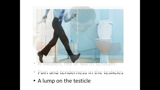
Epididymitis is infection or less frequently, inflammation of the epididymis (the coiled tube on the back of the testicle). The majority of men that develop epididymitis develop it because of a bacterial infection. Although males of any age can develop epididymitis, it occurs most frequently between ages of 20 to 39.
