
- Physical Examination
- Surgical Examination
- Ophthalmology
- Clinical Skills
- Orthopedics
- Surgery Videos
- Laparoscopy
- Pediatrics
- Funny Videos
- Cardiothoracic Surgery
- Nursing Videos
- Plastic Surgery
- Otorhinolaryngology
- Histology and Histopathology
- Neurosurgery
- Dermatology
- Pediatric Surgery
- Urology
- Dentistry
- Oncology and Cancers
- Anatomy Videos
- Health and Fitness
- Radiology
- Anaesthesia
- Physical Therapy
- Pharmacology
- Interventional Radiology
- Cardiology
- Endocrinology
- Gynecology
- Emergency Medicine
- Psychiatry and Psychology
- Childbirth Videos
- General Medical Videos
- Nephrology
- Physiology
- Diet and Food Health
- Diabetes Mellitus
- Neurology
- Women Health
- Osteoporosis
- Gastroenterology
- Pulmonology
- Hematology
- Rheumatology
- Toxicology
- Nuclear Medicine
- Infectious Diseases
- Vascular Disease
- Reproductive Health
- Burns and Wound Healing
- Other
Top videos


Dr. David Sneed of Aesthetica Med Spa in Austin discusses the latest liposuction technique known as Body Jet Water Liposuction - which is quickly gaining popularity due to the procedure being less invasive than traditional liposuction techniques, therefore minimizing recovery time and pain.
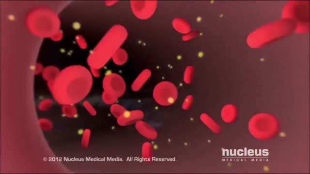
Diabetes Complications and Kidney Disease
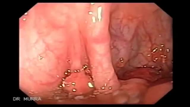
Juvenile polyposis syndrome (JPS) is a hereditary condition that is characterized by the presence of hamartomatous polyps in the digestive tract. Hamartomas are noncancerous (benign) masses of normal tissue that build up in the intestines or other places. These masses are called polyps if they develop inside a body structure, such as the intestines. The term “juvenile polyposis” refers to the type of polyp (juvenile polyp) that is found after examination of the polyp under a microscope, not the age at which people are diagnosed with JPS.

What happens when you wear High Heels. SHOW MORE
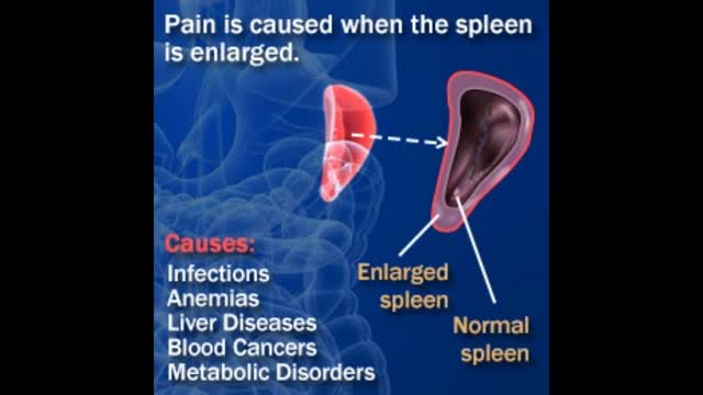
An enlarged spleen can be caused by infections, cirrhosis and other liver diseases, blood diseases characterized by abnormal blood cells, problems with the lymph system, or other conditions. Other causes of an enlarged spleen include: Inflammatory diseases such as sarcoidosis, lupus, and rheumatoid arthritis.

It's a symptom of heart disease but typically does not cause permanent damage to the heart. It is, though, a sign that you are a candidate for a heart attack at some point in the future. The chest pain may spread to your arm, shoulder, jaw, or back. It may feel like a pressure or squeezing sensation.
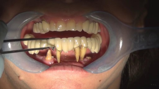
Watch that video of Removing Front Teeth
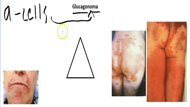
A glucagonoma is a rare tumor of the alpha cells of the pancreas that results in the overproduction of the hormone glucagon. Alpha cell tumors are commonly associated with glucagonoma syndrome, though similar symptoms are present in cases of pseudoglucagonoma syndrome in the absence of a glucagon-secreting tumor.

Laparoscopic anterior resection for cancer colon in Qatar by Dr. Al-Emadi

Womens Issues and Blood Clotting
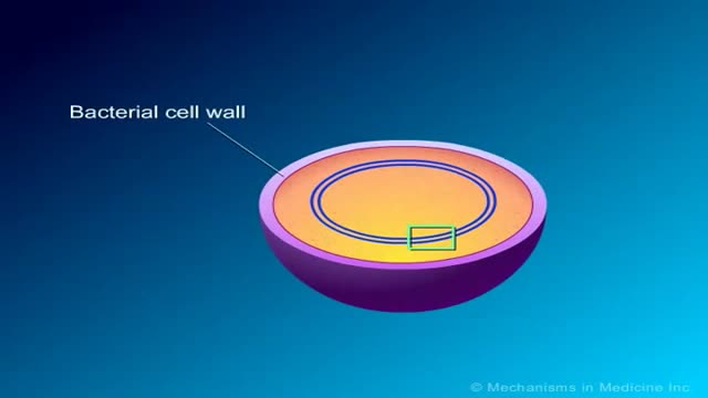
Macrolides Mechanisms of Action and Resistance

In the Womb - Identical Twins

Watch that video of 30 Pounds Of Poop Removed from Man's Stomach
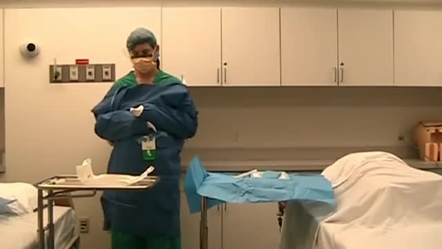
A central venous catheter (CVC), also known as a central line, central venous line, or central venous access catheter, is a catheter placed into a large vein. Catheters can be placed in veins in the neck (internal jugular vein), chest (subclavian vein or axillary vein), groin (femoral vein), or through veins in the arms (also known as a PICC line, or peripherally inserted central catheters). It is used to administer medication or fluids that are unable to be taken by mouth or would harm a smaller peripheral vein, obtain blood tests (specifically the "central venous oxygen saturation"), and measure central venous pressure.
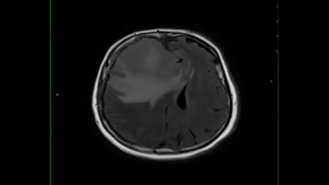
Lymphoma is a cancer that arises from the cells of the lymphatic system. In the brain, this type of cancer is called Primary CNS Lymphoma (PCNSL). Location. Lymphoma occurs most often in the cerebral hemisphere, but may also involve the cerebrospinal fluid, the eyes, or the spinal cord.

Eczema, or atopic dermatitis, is a rash that primarily occurs in people with asthma or allergies. The rash is often reddish and itchy with a scaly texture. Psoriasis is a common skin condition that can cause a scaly, itchy, red rash to form along the scalp, elbows, and joints.Apr 13, 2016
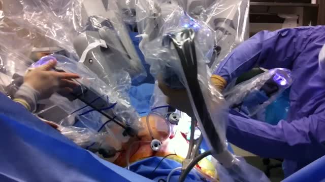
Endometrial cancer is a type of cancer that begins in the uterus. The uterus is the hollow, pear-shaped pelvic organ in women where fetal development occurs. Endometrial cancer begins in the layer of cells that form the lining (endometrium) of the uterus. Endometrial cancer is sometimes called uterine cancer. Other types of cancer can form in the uterus, including uterine sarcoma, but they are much less common than endometrial cancer. Endometrial cancer is often detected at an early stage because it frequently produces abnormal vaginal bleeding, which prompts women to see their doctors. If endometrial cancer is discovered early, removing the uterus surgically often cures endometrial cancer.
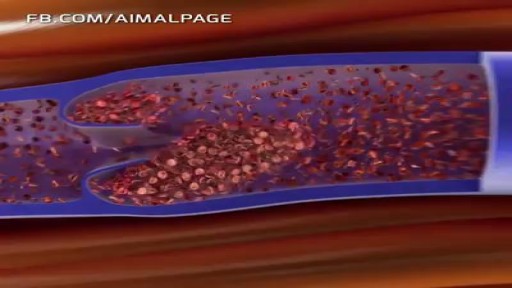
In Deep Vein Thrombosis, blood clot is able to reach the heart and from there it transport to the arteries of the lungs, where it may stuck in the...
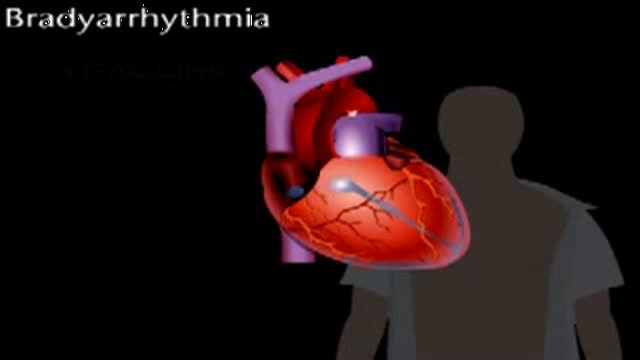
Bradyarrythmias
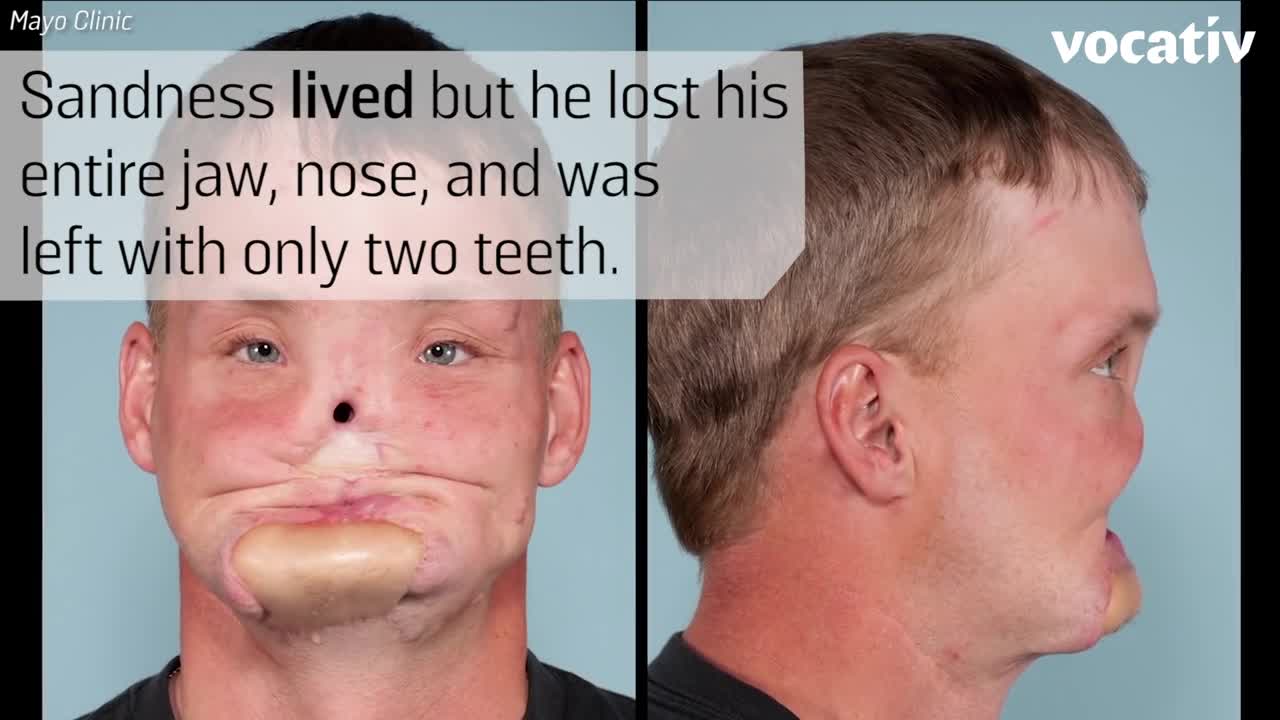
Doctors at the Mayo Clinic used 3D-printed models to prepare for their first-ever face transplant.
